Ali Salehi, Mohammad Malekahmadi, Abolfazl Karimi, Afsaneh Naderi Beni
{"title":"Retinal vascular changes after Silicon Oil removal in the Eye with Rhegmatogenous Retinal detachment.","authors":"Ali Salehi, Mohammad Malekahmadi, Abolfazl Karimi, Afsaneh Naderi Beni","doi":"10.1186/s40942-024-00587-9","DOIUrl":null,"url":null,"abstract":"<p><strong>Background: </strong>This study aims to examine vessel density changes in the optic nerve and macula following silicone oil removal (SOR) surgery in eyes with rhegmatogenous retinal detachment (RRD) at different time points by Optical Coherence Tomography Angiography (OCTA) in compared to the contralateral eye.</p><p><strong>Methods: </strong>A total of 43 eyes from 43 patients with silicone oil in their eyes for 3-9 months underwent OCT-A using AngioVue and optic disc-associated vessel density (VD) and thickness, macular-associated VD and thickness, Foveal avascular zone (FAZ) area, FAZ perimeter (PERIM), Acircularity index (AI), vessel density within a 300 μm wide region of the FAZ were compared between eyes. OCTA scans were performed one week before SOR and one month and three months after SOR.</p><p><strong>Results: </strong>The mean age of participants was 52.8 years (SD = 15.85) and a median visual acuity was 0.8 (range: 0.5-1.0). Notably, male participants constituted 67.4% of the sample. The preoperative mean value BCVA (logMAR) of patients was 0.73, and 3 months post-oil removal was 0.7727. Regarding optic disc parameters, RNFL thickness and vessel density (VD) measurements Peripapillary, whole disc, inside disc, and Disc Angio (superior, Nasal, inferior, temporal) did not change. In analyzing macular thickness parameters, all of them (Whole and Fovea, parafoveal, and Perifovea) remained unchanged. Examining macular vessel density parameters revealed no significant changes across superficial and deep retinal layers. Finally, the comparison of the foveal avascular zone (FAZ) area and flow density (FD) parameters demonstrated consistent measurements with non-significant alterations observed in FAZ size (p = 0.6) and FD values (p = 0.49) over the monitored duration.</p><p><strong>Conclusion: </strong>There was no change in peripapillary VD and macular vessel density of the superficial capillary plexus (SCP) and deep capillary plexus (DCP) after silicone oil removal. FAZ and full retinal thickness remained stable 3 month after SOR. Clinical trial number: Not applicable.</p>","PeriodicalId":14289,"journal":{"name":"International Journal of Retina and Vitreous","volume":"10 1","pages":"68"},"PeriodicalIF":2.4000,"publicationDate":"2024-09-30","publicationTypes":"Journal Article","fieldsOfStudy":null,"isOpenAccess":false,"openAccessPdf":"https://www.ncbi.nlm.nih.gov/pmc/articles/PMC11440900/pdf/","citationCount":"0","resultStr":null,"platform":"Semanticscholar","paperid":null,"PeriodicalName":"International Journal of Retina and Vitreous","FirstCategoryId":"1085","ListUrlMain":"https://doi.org/10.1186/s40942-024-00587-9","RegionNum":0,"RegionCategory":null,"ArticlePicture":[],"TitleCN":null,"AbstractTextCN":null,"PMCID":null,"EPubDate":"","PubModel":"","JCR":"Q2","JCRName":"OPHTHALMOLOGY","Score":null,"Total":0}
引用次数: 0
Abstract
Background: This study aims to examine vessel density changes in the optic nerve and macula following silicone oil removal (SOR) surgery in eyes with rhegmatogenous retinal detachment (RRD) at different time points by Optical Coherence Tomography Angiography (OCTA) in compared to the contralateral eye.
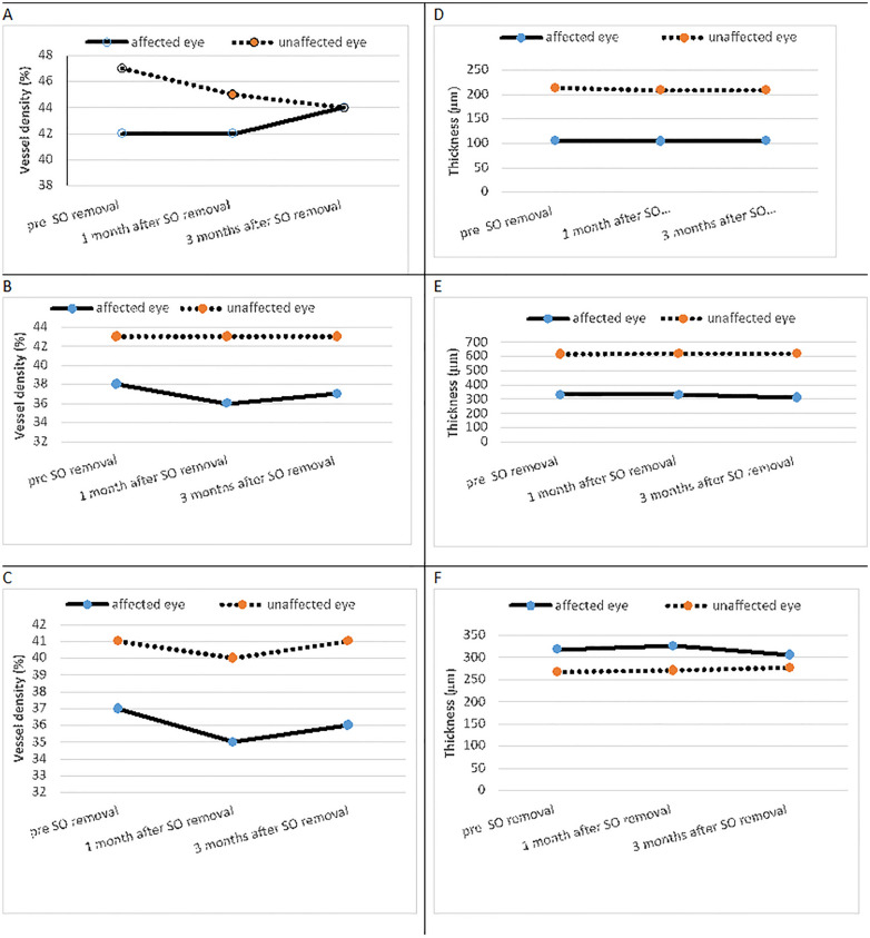
Methods: A total of 43 eyes from 43 patients with silicone oil in their eyes for 3-9 months underwent OCT-A using AngioVue and optic disc-associated vessel density (VD) and thickness, macular-associated VD and thickness, Foveal avascular zone (FAZ) area, FAZ perimeter (PERIM), Acircularity index (AI), vessel density within a 300 μm wide region of the FAZ were compared between eyes. OCTA scans were performed one week before SOR and one month and three months after SOR.
Results: The mean age of participants was 52.8 years (SD = 15.85) and a median visual acuity was 0.8 (range: 0.5-1.0). Notably, male participants constituted 67.4% of the sample. The preoperative mean value BCVA (logMAR) of patients was 0.73, and 3 months post-oil removal was 0.7727. Regarding optic disc parameters, RNFL thickness and vessel density (VD) measurements Peripapillary, whole disc, inside disc, and Disc Angio (superior, Nasal, inferior, temporal) did not change. In analyzing macular thickness parameters, all of them (Whole and Fovea, parafoveal, and Perifovea) remained unchanged. Examining macular vessel density parameters revealed no significant changes across superficial and deep retinal layers. Finally, the comparison of the foveal avascular zone (FAZ) area and flow density (FD) parameters demonstrated consistent measurements with non-significant alterations observed in FAZ size (p = 0.6) and FD values (p = 0.49) over the monitored duration.
Conclusion: There was no change in peripapillary VD and macular vessel density of the superficial capillary plexus (SCP) and deep capillary plexus (DCP) after silicone oil removal. FAZ and full retinal thickness remained stable 3 month after SOR. Clinical trial number: Not applicable.
期刊介绍:
International Journal of Retina and Vitreous focuses on the ophthalmic subspecialty of vitreoretinal disorders. The journal presents original articles on new approaches to diagnosis, outcomes of clinical trials, innovations in pharmacological therapy and surgical techniques, as well as basic science advances that impact clinical practice. Topical areas include, but are not limited to: -Imaging of the retina, choroid and vitreous -Innovations in optical coherence tomography (OCT) -Small-gauge vitrectomy, retinal detachment, chromovitrectomy -Electroretinography (ERG), microperimetry, other functional tests -Intraocular tumors -Retinal pharmacotherapy & drug delivery -Diabetic retinopathy & other vascular diseases -Age-related macular degeneration (AMD) & other macular entities


 分享
分享
 求助内容:
求助内容: 应助结果提醒方式:
 扫码关注我们
