Namra Ajmal, Yutao Deng, Lawrence C Kenyon, Mark T Curtis, Mauro Dispagna, Joseph Izes, Li Li
{"title":"Metastatic prostate adenocarcinoma to the brain - a clinicopathologic analysis of 21 cases.","authors":"Namra Ajmal, Yutao Deng, Lawrence C Kenyon, Mark T Curtis, Mauro Dispagna, Joseph Izes, Li Li","doi":"10.1186/s13000-024-01554-6","DOIUrl":null,"url":null,"abstract":"<p><strong>Background: </strong>Brain metastasis from prostate adenocarcinoma (PCa) is rare, often leading to death within a year. Its infrequent occurrence and atypical histopathologic features contribute to lower consideration in the differential diagnosis of tumor brain metastasis. This study aims to assess the clinical characteristics and distinctive histopathologic features of metastatic PCa in the brain for timely and enhanced diagnostic accuracy.</p><p><strong>Design: </strong>A retrospective search spanning 20 years (2003-2022) was conducted on our archives and identified 21 cases diagnosed as \"metastatic prostate adenocarcinoma (mPCa)\" in brain biopsies and resections. All existing slides were thoroughly reviewed to evaluate the histopathology of the mPCa.</p><p><strong>Result: </strong>The mean age at presentation for brain metastasis was 70 years. Of 21 cases, 5 were dural-based lesions, 16 were true intraparenchymal metastases, including 2 sellar/suprasellar masses, 3 frontal, 3 temporal, 3 occipital, 1 cerebellum, and 4 involving multiple brain lobes. The average interval between initial diagnosis and brain metastasis was 90.75 months. Notably, brain metastasis was the initial presentation for one patient, while another patient, initially diagnosed with prognostic grade group (GG) 2 PCa in 1/12 cores, presented with isolated brain metastasis two years later. Architecturally, tumor cells were arranged in sheets or nests in most cases; however, four cases showed histologic cribriform patterns, and five displayed papillary architecture. Cytohistology varied from uniform monomorphic to highly pleomorphic cells with prominent nucleoli (8/19) and high mitotic activity. Interestingly, 1 case showed small round blue cell morphology, another had focal areas of rhabdoid and spindle cell differentiation, and 6 had cytoplasmic clearing. Almost half of the cases (47%) showed necrosis.</p><p><strong>Conclusion: </strong>mPCa to the brain can present with variable histomorphology. Therefore, consideration of mPCa in the differential diagnosis of metastatic brain lesions, even with non-suggestive imaging, is imperative in male patients with or without a history of primary disease. Accurate and prompt diagnosis is crucial, given the recent advancements in treatment that have improved survival rates.</p>","PeriodicalId":11237,"journal":{"name":"Diagnostic Pathology","volume":"19 1","pages":"132"},"PeriodicalIF":2.3000,"publicationDate":"2024-10-01","publicationTypes":"Journal Article","fieldsOfStudy":null,"isOpenAccess":false,"openAccessPdf":"https://www.ncbi.nlm.nih.gov/pmc/articles/PMC11443825/pdf/","citationCount":"0","resultStr":null,"platform":"Semanticscholar","paperid":null,"PeriodicalName":"Diagnostic Pathology","FirstCategoryId":"3","ListUrlMain":"https://doi.org/10.1186/s13000-024-01554-6","RegionNum":3,"RegionCategory":"医学","ArticlePicture":[],"TitleCN":null,"AbstractTextCN":null,"PMCID":null,"EPubDate":"","PubModel":"","JCR":"Q2","JCRName":"PATHOLOGY","Score":null,"Total":0}
引用次数: 0
Abstract
Background: Brain metastasis from prostate adenocarcinoma (PCa) is rare, often leading to death within a year. Its infrequent occurrence and atypical histopathologic features contribute to lower consideration in the differential diagnosis of tumor brain metastasis. This study aims to assess the clinical characteristics and distinctive histopathologic features of metastatic PCa in the brain for timely and enhanced diagnostic accuracy.
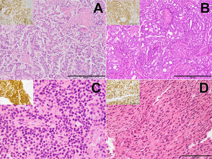
Design: A retrospective search spanning 20 years (2003-2022) was conducted on our archives and identified 21 cases diagnosed as "metastatic prostate adenocarcinoma (mPCa)" in brain biopsies and resections. All existing slides were thoroughly reviewed to evaluate the histopathology of the mPCa.
Result: The mean age at presentation for brain metastasis was 70 years. Of 21 cases, 5 were dural-based lesions, 16 were true intraparenchymal metastases, including 2 sellar/suprasellar masses, 3 frontal, 3 temporal, 3 occipital, 1 cerebellum, and 4 involving multiple brain lobes. The average interval between initial diagnosis and brain metastasis was 90.75 months. Notably, brain metastasis was the initial presentation for one patient, while another patient, initially diagnosed with prognostic grade group (GG) 2 PCa in 1/12 cores, presented with isolated brain metastasis two years later. Architecturally, tumor cells were arranged in sheets or nests in most cases; however, four cases showed histologic cribriform patterns, and five displayed papillary architecture. Cytohistology varied from uniform monomorphic to highly pleomorphic cells with prominent nucleoli (8/19) and high mitotic activity. Interestingly, 1 case showed small round blue cell morphology, another had focal areas of rhabdoid and spindle cell differentiation, and 6 had cytoplasmic clearing. Almost half of the cases (47%) showed necrosis.
Conclusion: mPCa to the brain can present with variable histomorphology. Therefore, consideration of mPCa in the differential diagnosis of metastatic brain lesions, even with non-suggestive imaging, is imperative in male patients with or without a history of primary disease. Accurate and prompt diagnosis is crucial, given the recent advancements in treatment that have improved survival rates.
期刊介绍:
Diagnostic Pathology is an open access, peer-reviewed, online journal that considers research in surgical and clinical pathology, immunology, and biology, with a special focus on cutting-edge approaches in diagnostic pathology and tissue-based therapy. The journal covers all aspects of surgical pathology, including classic diagnostic pathology, prognosis-related diagnosis (tumor stages, prognosis markers, such as MIB-percentage, hormone receptors, etc.), and therapy-related findings. The journal also focuses on the technological aspects of pathology, including molecular biology techniques, morphometry aspects (stereology, DNA analysis, syntactic structure analysis), communication aspects (telecommunication, virtual microscopy, virtual pathology institutions, etc.), and electronic education and quality assurance (for example interactive publication, on-line references with automated updating, etc.).


 分享
分享
 求助内容:
求助内容: 应助结果提醒方式:
 扫码关注我们
