Bo Ding, Biwei Mai, Tingyan Liu, Cuicui Liu, Hairong Bao, Jingzhou Hu, Xiaowen Qian, Song Wang, Qiuxiang Ou, Xiujuan Dong, Zhixian Lei, Gangfeng Yan
{"title":"Anlotinib treatment for rapidly progressing pediatric embryonal rhabdomyosarcoma in the maxillary gingiva: a case report.","authors":"Bo Ding, Biwei Mai, Tingyan Liu, Cuicui Liu, Hairong Bao, Jingzhou Hu, Xiaowen Qian, Song Wang, Qiuxiang Ou, Xiujuan Dong, Zhixian Lei, Gangfeng Yan","doi":"10.1186/s13000-024-01555-5","DOIUrl":null,"url":null,"abstract":"<p><strong>Background: </strong>Embryonal rhabdomyosarcoma (ERMS) is a highly aggressive form of soft-tissue sarcoma that predominantly affects children. Due to limited benefits and resistance to therapy, there is an unmet need to explore alternative therapeutic strategies.</p><p><strong>Case presentation: </strong>In this report, we present a rare case of pediatric ERMS located on the right side of the maxillary gingiva. A composite reference guide integrating clinical, radiographic, and histopathologic findings was used for a definitive diagnosis. Targeted next-generation sequencing of tumor biopsy was performed to identify genetic alterations. A 12-year-old female was admitted to the Pediatric Intensive Care Unit (PICU) and underwent a tracheotomy to relieve asphyxiation caused by a 5.5 cm diameter mass compressing the tongue root and pharyngeal cavity. Hematoxylin and eosin staining revealed a hybrid morphology characterized by clusters of round and spindle cells. Further immunohistochemistry assays indicated positive immunoreactivity for desmin, myogenin, and MyoD1. Various genetic alterations were identified, including mutations in GNAS, HRAS, LRP1B, amplification of MDM2 and IGF1R, and two novel IGF1R fusions. Negative PAX-FOXO1 fusion status supported the clinical diagnosis of ERMS. Initial treatment involved standard chemotherapy; however, the tumor persisted in its growth, reaching a maximum volume of 12 cm × 6 cm × 4 cm by the completion of treatment. Subsequent oral administration of anlotinib yielded a significant antitumor response, characterized by substantial tumor necrosis and size reduction. Following the ligation of the tumor pedicle and its removal, the patient developed a stabilized condition and was successfully discharged from PICU.</p><p><strong>Conclusions: </strong>Our study highlights the importance of accurate diagnosis established on multifaceted assessment for the effective treatment of ERMS. We present compelling evidence supporting the clinical use of anlotinib as a promising treatment strategy for pediatric ERMS patients, especially for those resistant to conventional chemotherapy.</p>","PeriodicalId":11237,"journal":{"name":"Diagnostic Pathology","volume":"19 1","pages":"135"},"PeriodicalIF":2.3000,"publicationDate":"2024-10-08","publicationTypes":"Journal Article","fieldsOfStudy":null,"isOpenAccess":false,"openAccessPdf":"https://www.ncbi.nlm.nih.gov/pmc/articles/PMC11460102/pdf/","citationCount":"0","resultStr":null,"platform":"Semanticscholar","paperid":null,"PeriodicalName":"Diagnostic Pathology","FirstCategoryId":"3","ListUrlMain":"https://doi.org/10.1186/s13000-024-01555-5","RegionNum":3,"RegionCategory":"医学","ArticlePicture":[],"TitleCN":null,"AbstractTextCN":null,"PMCID":null,"EPubDate":"","PubModel":"","JCR":"Q2","JCRName":"PATHOLOGY","Score":null,"Total":0}
引用次数: 0
Abstract
Background: Embryonal rhabdomyosarcoma (ERMS) is a highly aggressive form of soft-tissue sarcoma that predominantly affects children. Due to limited benefits and resistance to therapy, there is an unmet need to explore alternative therapeutic strategies.
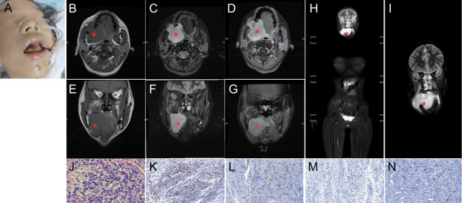
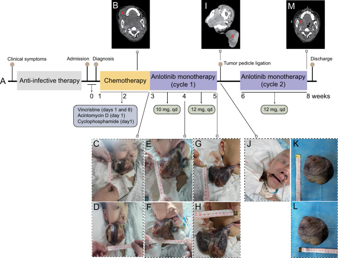
Case presentation: In this report, we present a rare case of pediatric ERMS located on the right side of the maxillary gingiva. A composite reference guide integrating clinical, radiographic, and histopathologic findings was used for a definitive diagnosis. Targeted next-generation sequencing of tumor biopsy was performed to identify genetic alterations. A 12-year-old female was admitted to the Pediatric Intensive Care Unit (PICU) and underwent a tracheotomy to relieve asphyxiation caused by a 5.5 cm diameter mass compressing the tongue root and pharyngeal cavity. Hematoxylin and eosin staining revealed a hybrid morphology characterized by clusters of round and spindle cells. Further immunohistochemistry assays indicated positive immunoreactivity for desmin, myogenin, and MyoD1. Various genetic alterations were identified, including mutations in GNAS, HRAS, LRP1B, amplification of MDM2 and IGF1R, and two novel IGF1R fusions. Negative PAX-FOXO1 fusion status supported the clinical diagnosis of ERMS. Initial treatment involved standard chemotherapy; however, the tumor persisted in its growth, reaching a maximum volume of 12 cm × 6 cm × 4 cm by the completion of treatment. Subsequent oral administration of anlotinib yielded a significant antitumor response, characterized by substantial tumor necrosis and size reduction. Following the ligation of the tumor pedicle and its removal, the patient developed a stabilized condition and was successfully discharged from PICU.
Conclusions: Our study highlights the importance of accurate diagnosis established on multifaceted assessment for the effective treatment of ERMS. We present compelling evidence supporting the clinical use of anlotinib as a promising treatment strategy for pediatric ERMS patients, especially for those resistant to conventional chemotherapy.
期刊介绍:
Diagnostic Pathology is an open access, peer-reviewed, online journal that considers research in surgical and clinical pathology, immunology, and biology, with a special focus on cutting-edge approaches in diagnostic pathology and tissue-based therapy. The journal covers all aspects of surgical pathology, including classic diagnostic pathology, prognosis-related diagnosis (tumor stages, prognosis markers, such as MIB-percentage, hormone receptors, etc.), and therapy-related findings. The journal also focuses on the technological aspects of pathology, including molecular biology techniques, morphometry aspects (stereology, DNA analysis, syntactic structure analysis), communication aspects (telecommunication, virtual microscopy, virtual pathology institutions, etc.), and electronic education and quality assurance (for example interactive publication, on-line references with automated updating, etc.).


 分享
分享
 求助内容:
求助内容: 应助结果提醒方式:
 扫码关注我们
