Proteomic Analysis Reveals Major Proteins and Pathways That Mediate the Effect of 17-β-Estradiol in Cell Division and Apoptosis in Breast Cancer MCF7 Cells.
Zhenqi Zhou, Brihget Sicairos, Jianhong Zhou, Yuchun Du
{"title":"Proteomic Analysis Reveals Major Proteins and Pathways That Mediate the Effect of 17-β-Estradiol in Cell Division and Apoptosis in Breast Cancer MCF7 Cells.","authors":"Zhenqi Zhou, Brihget Sicairos, Jianhong Zhou, Yuchun Du","doi":"10.1021/acs.jproteome.4c00102","DOIUrl":null,"url":null,"abstract":"<p><p>Despite extensive research, the genes/proteins and pathways responsible for the physiological effects of estrogen remain elusive. In this study, we determined the effect of estrogen on global protein expression in breast cancer MCF7 cells using a proteomic method. The expression of 77 cytosolic, 74 nuclear, and 81 membrane/organelle proteins was significantly altered by 17-β-estradiol (E2). Protein enrichment analyses suggest that E2 may stimulate cell division primarily by promoting the G1 to S phase transition and advancing the G2/M checkpoint. The effect of E2 on cell survival was complex, as it could simultaneously enhance and inhibit apoptosis. Bioinformatics analysis suggests that E2 may enhance apoptosis by promoting the accumulation of the pore-forming protein Bax in the mitochondria and inhibit apoptosis by activating the PI3K/AKT/mTOR signaling pathway. We verified the activation of the PI3K signaling and the accumulation of Bax in the membrane/organelle fraction in E2-treated cells using immunoblotting. Treatment of MCF7 cells with E2 and the PI3K inhibitor Ly294002 significantly enhanced apoptosis compared to those treated with E2 alone, suggesting that combining estrogen with a PI3K inhibitor could be a promising strategy for treating ERα-positive breast cancer. Interestingly, many of the E2-upregulated proteins contained the HEAT, KH, and RRM domains.</p>","PeriodicalId":48,"journal":{"name":"Journal of Proteome Research","volume":" ","pages":"4835-4848"},"PeriodicalIF":3.6000,"publicationDate":"2024-11-01","publicationTypes":"Journal Article","fieldsOfStudy":null,"isOpenAccess":false,"openAccessPdf":"https://www.ncbi.nlm.nih.gov/pmc/articles/PMC11536429/pdf/","citationCount":"0","resultStr":null,"platform":"Semanticscholar","paperid":null,"PeriodicalName":"Journal of Proteome Research","FirstCategoryId":"99","ListUrlMain":"https://doi.org/10.1021/acs.jproteome.4c00102","RegionNum":2,"RegionCategory":"生物学","ArticlePicture":[],"TitleCN":null,"AbstractTextCN":null,"PMCID":null,"EPubDate":"2024/10/11 0:00:00","PubModel":"Epub","JCR":"Q1","JCRName":"BIOCHEMICAL RESEARCH METHODS","Score":null,"Total":0}
引用次数: 0
Abstract
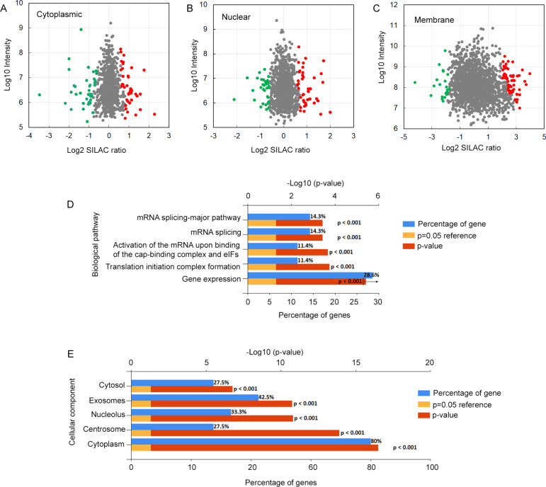
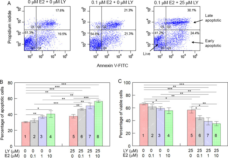
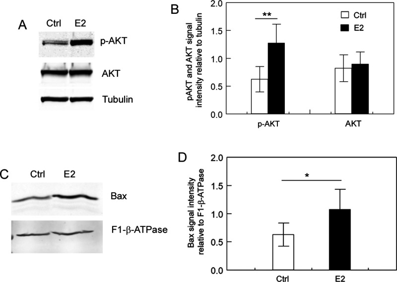
Despite extensive research, the genes/proteins and pathways responsible for the physiological effects of estrogen remain elusive. In this study, we determined the effect of estrogen on global protein expression in breast cancer MCF7 cells using a proteomic method. The expression of 77 cytosolic, 74 nuclear, and 81 membrane/organelle proteins was significantly altered by 17-β-estradiol (E2). Protein enrichment analyses suggest that E2 may stimulate cell division primarily by promoting the G1 to S phase transition and advancing the G2/M checkpoint. The effect of E2 on cell survival was complex, as it could simultaneously enhance and inhibit apoptosis. Bioinformatics analysis suggests that E2 may enhance apoptosis by promoting the accumulation of the pore-forming protein Bax in the mitochondria and inhibit apoptosis by activating the PI3K/AKT/mTOR signaling pathway. We verified the activation of the PI3K signaling and the accumulation of Bax in the membrane/organelle fraction in E2-treated cells using immunoblotting. Treatment of MCF7 cells with E2 and the PI3K inhibitor Ly294002 significantly enhanced apoptosis compared to those treated with E2 alone, suggesting that combining estrogen with a PI3K inhibitor could be a promising strategy for treating ERα-positive breast cancer. Interestingly, many of the E2-upregulated proteins contained the HEAT, KH, and RRM domains.
期刊介绍:
Journal of Proteome Research publishes content encompassing all aspects of global protein analysis and function, including the dynamic aspects of genomics, spatio-temporal proteomics, metabonomics and metabolomics, clinical and agricultural proteomics, as well as advances in methodology including bioinformatics. The theme and emphasis is on a multidisciplinary approach to the life sciences through the synergy between the different types of "omics".


 分享
分享
 求助内容:
求助内容: 应助结果提醒方式:
 扫码关注我们
