A novel method to detect carotid artery in-stent restenosis

IF 2.3 4区 医学 Q3 CLINICAL NEUROLOGY Journal of Neuroimaging Pub Date : 2024-10-27 DOI:10.1111/jon.13245
Yuding Luo, Gangfeng Gu, Yan Li, Bo Zheng, Fanzhou Ren, Junqiu Wang, Chuanli Chen, Zhao Chen, Yingqian Zhang, Bangcheng Zhao, Jian Yang, Jian Wang
{"title":"A novel method to detect carotid artery in-stent restenosis","authors":"Yuding Luo,&nbsp;Gangfeng Gu,&nbsp;Yan Li,&nbsp;Bo Zheng,&nbsp;Fanzhou Ren,&nbsp;Junqiu Wang,&nbsp;Chuanli Chen,&nbsp;Zhao Chen,&nbsp;Yingqian Zhang,&nbsp;Bangcheng Zhao,&nbsp;Jian Yang,&nbsp;Jian Wang","doi":"10.1111/jon.13245","DOIUrl":null,"url":null,"abstract":"<div>\n \n \n <section>\n \n <h3> Background and Purpose</h3>\n \n <p>Carotid artery stenosis is a major risk factor for ischemic stroke. Despite carotid artery stenting, in-stent restenosis (ISR) remains challenging. Pigs serve as an ideal ISR model. This study aims to establish a novel porcine model of carotid ISR using open-loop and closed-loop stents and to assess ISR with optical coherence tomography (OCT) and histopathology, comparing incidence and vascular response between stent types.</p>\n </section>\n \n <section>\n \n <h3> Methods</h3>\n \n <p>Twelve adult male Bama miniature pigs underwent carotid stenting with either open-loop or closed-loop stents. The animals received antiplatelet therapy pre- and postimplantation. Postimplantation evaluations at 90 days included carotid digital subtraction angiography (DSA), OCT, histopathological examination, and electron microscopy.</p>\n </section>\n \n <section>\n \n <h3> Results</h3>\n \n <p>Both stent types showed ISR as detected by OCT and DSA. OCT revealed comparable neointimal proliferation within stent struts for both types, with no significant differences in stent, lumen, and neointimal dimensions. Histopathological analysis and electron microscopy provided insights into tissue responses and healing processes following stent implantation. No significant difference in ISR incidence was found between the stent types based on a <i>χ</i><sup>2</sup> test (<i>p</i> = .110). OCT and hematoxylin-eosin staining exhibit the highest consistency in evaluating neointimal area.</p>\n </section>\n \n <section>\n \n <h3> Conclusions</h3>\n \n <p>The novel porcine ISR model demonstrated similar ISR outcomes for open-loop and closed-loop stents. OCT proved to be a highly consistent and valuable tool for evaluating stent and arterial conditions, comparable to histopathological findings. However, due to a small sample size, the validity of these preliminary findings requires further investigation to be confirmed.</p>\n </section>\n </div>","PeriodicalId":16399,"journal":{"name":"Journal of Neuroimaging","volume":"34 6","pages":"664-672"},"PeriodicalIF":2.3000,"publicationDate":"2024-10-27","publicationTypes":"Journal Article","fieldsOfStudy":null,"isOpenAccess":false,"openAccessPdf":"https://onlinelibrary.wiley.com/doi/epdf/10.1111/jon.13245","citationCount":"0","resultStr":null,"platform":"Semanticscholar","paperid":null,"PeriodicalName":"Journal of Neuroimaging","FirstCategoryId":"3","ListUrlMain":"https://onlinelibrary.wiley.com/doi/10.1111/jon.13245","RegionNum":4,"RegionCategory":"医学","ArticlePicture":[],"TitleCN":null,"AbstractTextCN":null,"PMCID":null,"EPubDate":"","PubModel":"","JCR":"Q3","JCRName":"CLINICAL NEUROLOGY","Score":null,"Total":0}
引用次数: 0

Abstract

Background and Purpose

Carotid artery stenosis is a major risk factor for ischemic stroke. Despite carotid artery stenting, in-stent restenosis (ISR) remains challenging. Pigs serve as an ideal ISR model. This study aims to establish a novel porcine model of carotid ISR using open-loop and closed-loop stents and to assess ISR with optical coherence tomography (OCT) and histopathology, comparing incidence and vascular response between stent types.

Methods

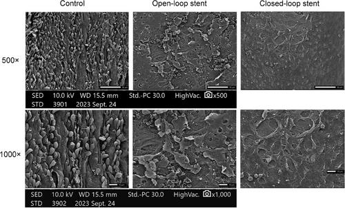
Twelve adult male Bama miniature pigs underwent carotid stenting with either open-loop or closed-loop stents. The animals received antiplatelet therapy pre- and postimplantation. Postimplantation evaluations at 90 days included carotid digital subtraction angiography (DSA), OCT, histopathological examination, and electron microscopy.

Results

Both stent types showed ISR as detected by OCT and DSA. OCT revealed comparable neointimal proliferation within stent struts for both types, with no significant differences in stent, lumen, and neointimal dimensions. Histopathological analysis and electron microscopy provided insights into tissue responses and healing processes following stent implantation. No significant difference in ISR incidence was found between the stent types based on a χ2 test (p = .110). OCT and hematoxylin-eosin staining exhibit the highest consistency in evaluating neointimal area.

Conclusions

The novel porcine ISR model demonstrated similar ISR outcomes for open-loop and closed-loop stents. OCT proved to be a highly consistent and valuable tool for evaluating stent and arterial conditions, comparable to histopathological findings. However, due to a small sample size, the validity of these preliminary findings requires further investigation to be confirmed.
Abstract Image

查看原文
分享 分享
微信好友 朋友圈 QQ好友 复制链接
本刊更多论文
检测颈动脉支架内再狭窄的新方法
背景和目的:颈动脉狭窄是缺血性中风的主要风险因素。尽管进行了颈动脉支架植入术,但支架内再狭窄(ISR)仍然是一个难题。猪是理想的 ISR 模型。本研究旨在使用开环和闭环支架建立新型颈动脉ISR猪模型,并通过光学相干断层扫描(OCT)和组织病理学评估ISR,比较不同类型支架的发病率和血管反应:12头成年雄性巴马微型猪接受了颈动脉支架手术,支架分为开环支架和闭环支架。这些动物在植入前后均接受了抗血小板治疗。植入后90天的评估包括颈动脉数字减影血管造影(DSA)、OCT、组织病理学检查和电子显微镜检查:结果:OCT和DSA检测到两种类型的支架都出现了ISR。OCT显示两种类型的支架支柱内都有类似的新内膜增生,支架、管腔和新内膜尺寸无明显差异。组织病理学分析和电子显微镜检查有助于了解支架植入后的组织反应和愈合过程。根据χ2检验(P = .110),两种支架类型的ISR发生率无明显差异。OCT 和苏木精-伊红染色在评估新生内膜面积方面表现出最高的一致性:结论:新型猪 ISR 模型显示,开环和闭环支架的 ISR 结果相似。OCT 被证明是评估支架和动脉状况的高度一致且有价值的工具,可与组织病理学结果相媲美。然而,由于样本量较小,这些初步研究结果的有效性还需要进一步的研究来证实。
本文章由计算机程序翻译,如有差异,请以英文原文为准。
求助全文
约1分钟内获得全文 去求助
来源期刊
Journal of Neuroimaging
Journal of Neuroimaging 医学-核医学
CiteScore
4.70
自引率
0.00%
发文量
117
审稿时长
6-12 weeks
期刊介绍: Start reading the Journal of Neuroimaging to learn the latest neurological imaging techniques. The peer-reviewed research is written in a practical clinical context, giving you the information you need on: MRI CT Carotid Ultrasound and TCD SPECT PET Endovascular Surgical Neuroradiology Functional MRI Xenon CT and other new and upcoming neuroscientific modalities.The Journal of Neuroimaging addresses the full spectrum of human nervous system disease, including stroke, neoplasia, degenerating and demyelinating disease, epilepsy, tumors, lesions, infectious disease, cerebral vascular arterial diseases, toxic-metabolic disease, psychoses, dementias, heredo-familial disease, and trauma.Offering original research, review articles, case reports, neuroimaging CPCs, and evaluations of instruments and technology relevant to the nervous system, the Journal of Neuroimaging focuses on useful clinical developments and applications, tested techniques and interpretations, patient care, diagnostics, and therapeutics. Start reading today!
期刊最新文献
Predicting glioblastoma progression using MR diffusion tensor imaging: A systematic review Nerve cross-sectional area in vincristine-induced polyneuropathy: A nerve ultrasound pilot study Comparison of antithrombogenic coated and uncoated flow diverters in ruptured and unruptured cerebral aneurysms Functional MRI and cognition in multiple sclerosis—Where are we now? Time-dependent MR diffusion analysis of functioning and nonfunctioning pituitary adenomas/pituitary neuroendocrine tumors
×
引用
GB/T 7714-2015
复制
MLA
复制
APA
复制
导出至
BibTeX EndNote RefMan NoteFirst NoteExpress
×
×
提示
您的信息不完整,为了账户安全,请先补充。
现在去补充
×
提示
您因"违规操作"
具体请查看互助需知
我知道了
×
提示
现在去查看 取消
×
提示
确定
0
微信
客服QQ
Book学术公众号 扫码关注我们
反馈
×
意见反馈
请填写您的意见或建议
请填写您的手机或邮箱
已复制链接
已复制链接
快去分享给好友吧!
我知道了
×
扫码分享
扫码分享
Book学术官方微信
Book学术文献互助
Book学术文献互助群
群 号:481959085
Book学术
文献互助 智能选刊 最新文献 互助须知 联系我们:info@booksci.cn
Book学术提供免费学术资源搜索服务,方便国内外学者检索中英文文献。致力于提供最便捷和优质的服务体验。
Copyright © 2023 Book学术 All rights reserved.
ghs 京公网安备 11010802042870号 京ICP备2023020795号-1