Jan-Hannes Schäfer, Lena Clausmeyer, Carolin Körner, Bianca M. Esch, Verena N. Wolf, Jennifer Sapia, Yara Ahmed, Stefan Walter, Stefano Vanni, Dovile Januliene, Arne Moeller, Florian Fröhlich
{"title":"Structure of the yeast ceramide synthase","authors":"Jan-Hannes Schäfer, Lena Clausmeyer, Carolin Körner, Bianca M. Esch, Verena N. Wolf, Jennifer Sapia, Yara Ahmed, Stefan Walter, Stefano Vanni, Dovile Januliene, Arne Moeller, Florian Fröhlich","doi":"10.1038/s41594-024-01415-2","DOIUrl":null,"url":null,"abstract":"Ceramides are essential lipids involved in forming complex sphingolipids and acting as signaling molecules. They result from the N-acylation of a sphingoid base and a CoA-activated fatty acid, a reaction catalyzed by the ceramide synthase (CerS) family of enzymes. Yet, the precise structural details and catalytic mechanisms of CerSs have remained elusive. Here we used cryo-electron microscopy single-particle analysis to unravel the structure of the yeast CerS complex in both an active and a fumonisin B1-inhibited state. Our results reveal the complex’s architecture as a dimer of Lip1 subunits bound to the catalytic subunits Lag1 and Lac1. Each catalytic subunit forms a hydrophobic crevice connecting the cytosolic site with the intermembrane space. The active site, located centrally in the tunnel, was resolved in a substrate preloaded state, representing one intermediate in ceramide synthesis. Our data provide evidence for competitive binding of fumonisin B1 to the acyl-CoA-binding tunnel. Using cryo-electron microscopy, Schäfer et al. solved the structure of the yeast ceramide synthase complex, consisting of Lip1, Lag1 and Lac1 subunits. They found that fumonisin B1 binds competitively at a key site, suggesting a mechanism for ceramide synthesis.","PeriodicalId":49141,"journal":{"name":"Nature Structural & Molecular Biology","volume":"32 3","pages":"441-449"},"PeriodicalIF":10.1000,"publicationDate":"2024-11-11","publicationTypes":"Journal Article","fieldsOfStudy":null,"isOpenAccess":false,"openAccessPdf":"","citationCount":"0","resultStr":null,"platform":"Semanticscholar","paperid":null,"PeriodicalName":"Nature Structural & Molecular Biology","FirstCategoryId":"99","ListUrlMain":"https://www.nature.com/articles/s41594-024-01415-2","RegionNum":1,"RegionCategory":"生物学","ArticlePicture":[],"TitleCN":null,"AbstractTextCN":null,"PMCID":null,"EPubDate":"","PubModel":"","JCR":"Q1","JCRName":"BIOCHEMISTRY & MOLECULAR BIOLOGY","Score":null,"Total":0}
引用次数: 0
Abstract
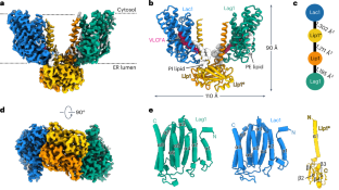
Ceramides are essential lipids involved in forming complex sphingolipids and acting as signaling molecules. They result from the N-acylation of a sphingoid base and a CoA-activated fatty acid, a reaction catalyzed by the ceramide synthase (CerS) family of enzymes. Yet, the precise structural details and catalytic mechanisms of CerSs have remained elusive. Here we used cryo-electron microscopy single-particle analysis to unravel the structure of the yeast CerS complex in both an active and a fumonisin B1-inhibited state. Our results reveal the complex’s architecture as a dimer of Lip1 subunits bound to the catalytic subunits Lag1 and Lac1. Each catalytic subunit forms a hydrophobic crevice connecting the cytosolic site with the intermembrane space. The active site, located centrally in the tunnel, was resolved in a substrate preloaded state, representing one intermediate in ceramide synthesis. Our data provide evidence for competitive binding of fumonisin B1 to the acyl-CoA-binding tunnel. Using cryo-electron microscopy, Schäfer et al. solved the structure of the yeast ceramide synthase complex, consisting of Lip1, Lag1 and Lac1 subunits. They found that fumonisin B1 binds competitively at a key site, suggesting a mechanism for ceramide synthesis.
期刊介绍:
Nature Structural & Molecular Biology is a comprehensive platform that combines structural and molecular research. Our journal focuses on exploring the functional and mechanistic aspects of biological processes, emphasizing how molecular components collaborate to achieve a particular function. While structural data can shed light on these insights, our publication does not require them as a prerequisite.


 分享
分享
 求助内容:
求助内容: 应助结果提醒方式:
 扫码关注我们
