Nicole Wake, Yenpo Lin, Ek T Tan, Darryl B Sneag, Sarah Ianucci, Maggie Fung
{"title":"3D printing of the brachial plexus and its osseous landmarks using magnetic resonance neurography for thoracic outlet syndrome evaluation.","authors":"Nicole Wake, Yenpo Lin, Ek T Tan, Darryl B Sneag, Sarah Ianucci, Maggie Fung","doi":"10.1186/s41205-024-00239-6","DOIUrl":null,"url":null,"abstract":"<p><strong>Background: </strong>Patient-specific three-dimensional (3D) printed anatomic models are valuable clinical tools that facilitate enhanced visualization of pertinent anatomic structures and have demonstrated benefits of reduced surgical times, increased surgeon confidence, and improved operative results and subsequent patient outcomes. Medical image-based 3D printed anatomic models are generally created from computed tomography (CT), however magnetic resonance imaging (MRI), which offers exquisite soft tissue characterization and flexible contrast avoiding the use of ionizing radiation, is an attractive alternative. Herein, the application of 3D printing incorporating both MR neurography and zero-echo time (ZTE) MRI for visualization of the brachial plexus anatomy in a subject with thoracic outlet syndrome (TOS) is described.</p><p><strong>Methods: </strong>A 28-year-old man presented with chronic right upper limb discomfort and paresthesias extending from the shoulder region to the third and fourth digits. The subject underwent evaluation with a unilateral brachial plexus MR neurography protocol at 3.0 Tesla for suspicion of TOS. The protocol included T2-weighted, 3D fast spin echo short-tau inversion recovery (STIR-FSE) and 3D radial ZTE sequences for depiction of the nerves and bones, respectively. The first rib and its synostosis impinged upon the inferior aspect of the T1 nerve root (T1NR), with accompanying mild enlargement of the T1NR. A 3D printed anatomic model was created and included: (1) bone (spine, ribs, clavicle, scapula, and humerus), (2) brachial plexus, and (3) costal cartilage.</p><p><strong>Results: </strong>The 3D printed model clearly demonstrated a T1NR impingement from the synostosis, confirming the diagnosis of neurologic thoracic outlet syndrome (TOS) and guided the treatment approach in prescribing TOS-specific physical therapy, which led to significant improvements in the patient's condition.</p><p><strong>Conclusion: </strong>To our knowledge, this is the first in-vivo human 3D printed case for TOS using MRI-only data. The 3D printed model allowed for improved visualization and understanding of the spatial relationships between the nerves of the brachial plexus and surrounding osseous structures responsible for the patient's symptoms.</p><p><strong>Clinical trial number: </strong>Not applicable.</p>","PeriodicalId":72036,"journal":{"name":"3D printing in medicine","volume":"10 1","pages":"36"},"PeriodicalIF":3.1000,"publicationDate":"2024-11-14","publicationTypes":"Journal Article","fieldsOfStudy":null,"isOpenAccess":false,"openAccessPdf":"https://www.ncbi.nlm.nih.gov/pmc/articles/PMC11562346/pdf/","citationCount":"0","resultStr":null,"platform":"Semanticscholar","paperid":null,"PeriodicalName":"3D printing in medicine","FirstCategoryId":"1085","ListUrlMain":"https://doi.org/10.1186/s41205-024-00239-6","RegionNum":0,"RegionCategory":null,"ArticlePicture":[],"TitleCN":null,"AbstractTextCN":null,"PMCID":null,"EPubDate":"","PubModel":"","JCR":"Q1","JCRName":"RADIOLOGY, NUCLEAR MEDICINE & MEDICAL IMAGING","Score":null,"Total":0}
引用次数: 0
Abstract
Background: Patient-specific three-dimensional (3D) printed anatomic models are valuable clinical tools that facilitate enhanced visualization of pertinent anatomic structures and have demonstrated benefits of reduced surgical times, increased surgeon confidence, and improved operative results and subsequent patient outcomes. Medical image-based 3D printed anatomic models are generally created from computed tomography (CT), however magnetic resonance imaging (MRI), which offers exquisite soft tissue characterization and flexible contrast avoiding the use of ionizing radiation, is an attractive alternative. Herein, the application of 3D printing incorporating both MR neurography and zero-echo time (ZTE) MRI for visualization of the brachial plexus anatomy in a subject with thoracic outlet syndrome (TOS) is described.
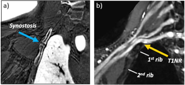
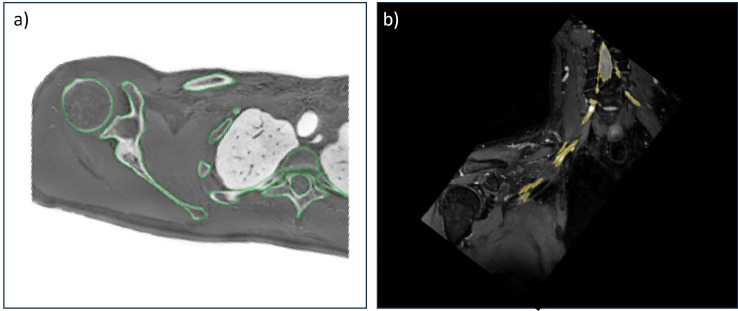
Methods: A 28-year-old man presented with chronic right upper limb discomfort and paresthesias extending from the shoulder region to the third and fourth digits. The subject underwent evaluation with a unilateral brachial plexus MR neurography protocol at 3.0 Tesla for suspicion of TOS. The protocol included T2-weighted, 3D fast spin echo short-tau inversion recovery (STIR-FSE) and 3D radial ZTE sequences for depiction of the nerves and bones, respectively. The first rib and its synostosis impinged upon the inferior aspect of the T1 nerve root (T1NR), with accompanying mild enlargement of the T1NR. A 3D printed anatomic model was created and included: (1) bone (spine, ribs, clavicle, scapula, and humerus), (2) brachial plexus, and (3) costal cartilage.
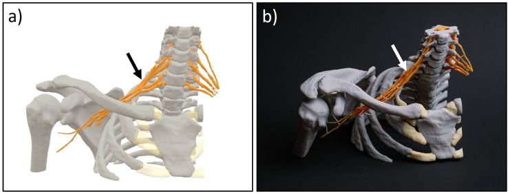
Results: The 3D printed model clearly demonstrated a T1NR impingement from the synostosis, confirming the diagnosis of neurologic thoracic outlet syndrome (TOS) and guided the treatment approach in prescribing TOS-specific physical therapy, which led to significant improvements in the patient's condition.
Conclusion: To our knowledge, this is the first in-vivo human 3D printed case for TOS using MRI-only data. The 3D printed model allowed for improved visualization and understanding of the spatial relationships between the nerves of the brachial plexus and surrounding osseous structures responsible for the patient's symptoms.


 分享
分享

 求助内容:
求助内容: 应助结果提醒方式:
 扫码关注我们
