{"title":"Deep Learning Algorithm for Keratoconus Detection from Tomographic Maps and Corneal Biomechanics: A Diagnostic Study.","authors":"Wiyada Quanchareonsap, Ngamjit Kasetsuwan, Usanee Reinprayoon, Yonrawee Piyacomn, Thitima Wungcharoen, Monthira Jermjutitham","doi":"10.4103/joco.joco_18_24","DOIUrl":null,"url":null,"abstract":"<p><strong>Purpose: </strong>To develop an artificial intelligence (AI) approach for differentiating between normal cornea, subclinical, and keratoconus (KC) using tomographic maps from Pentacam (Oculus) and corneal biomechanics from Corvis ST (Oculus).</p><p><strong>Methods: </strong>A total of 1,668 tomographic (769 patients) and 611 biomechanical (307 patients) images from the Chula Refractive Surgery Center, King Chulalongkorn Memorial Hospital were included. The sample size was divided into the Pentacam and combined Pentacam-Corvis groups. Different convolutional neural network approaches were used to enhance the KC and subclinical KC detection performance.</p><p><strong>Results: </strong>AI model 1, which obtained refractive maps from Pentacam, achieved an area under the receiver operating characteristic curve (AUC) of 0.938 and accuracy of 0.947 (sensitivity, 90.8% and specificity, 96.9%). AI model 2, which added dynamic corneal response and the Vinciguerra screening report from Corvis ST to AI Model 1, achieved an AUC of 0.985 and accuracy of 0.956 (sensitivity, 93.0% and specificity, 94.3%). AI model 3, which added the corneal biomechanical index to AI Model 2, reached an AUC of 0.991 and accuracy of 0.956 (sensitivity, 93.0% and specificity, 94.3%).</p><p><strong>Conclusions: </strong>Our study showed that AI models using either anterior corneal curvature alone or combined with corneal biomechanics could help classify normal and keratoconic corneas, which would make diagnosis more accurate and would be helpful in decision-making for the treatment.</p>","PeriodicalId":15423,"journal":{"name":"Journal of Current Ophthalmology","volume":"36 1","pages":"46-53"},"PeriodicalIF":0.9000,"publicationDate":"2024-10-16","publicationTypes":"Journal Article","fieldsOfStudy":null,"isOpenAccess":false,"openAccessPdf":"https://www.ncbi.nlm.nih.gov/pmc/articles/PMC11567606/pdf/","citationCount":"0","resultStr":null,"platform":"Semanticscholar","paperid":null,"PeriodicalName":"Journal of Current Ophthalmology","FirstCategoryId":"1085","ListUrlMain":"https://doi.org/10.4103/joco.joco_18_24","RegionNum":0,"RegionCategory":null,"ArticlePicture":[],"TitleCN":null,"AbstractTextCN":null,"PMCID":null,"EPubDate":"2024/1/1 0:00:00","PubModel":"eCollection","JCR":"Q3","JCRName":"OPHTHALMOLOGY","Score":null,"Total":0}
引用次数: 0
Abstract
Purpose: To develop an artificial intelligence (AI) approach for differentiating between normal cornea, subclinical, and keratoconus (KC) using tomographic maps from Pentacam (Oculus) and corneal biomechanics from Corvis ST (Oculus).
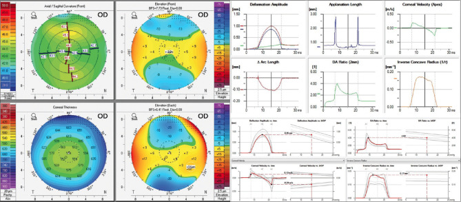
Methods: A total of 1,668 tomographic (769 patients) and 611 biomechanical (307 patients) images from the Chula Refractive Surgery Center, King Chulalongkorn Memorial Hospital were included. The sample size was divided into the Pentacam and combined Pentacam-Corvis groups. Different convolutional neural network approaches were used to enhance the KC and subclinical KC detection performance.
Results: AI model 1, which obtained refractive maps from Pentacam, achieved an area under the receiver operating characteristic curve (AUC) of 0.938 and accuracy of 0.947 (sensitivity, 90.8% and specificity, 96.9%). AI model 2, which added dynamic corneal response and the Vinciguerra screening report from Corvis ST to AI Model 1, achieved an AUC of 0.985 and accuracy of 0.956 (sensitivity, 93.0% and specificity, 94.3%). AI model 3, which added the corneal biomechanical index to AI Model 2, reached an AUC of 0.991 and accuracy of 0.956 (sensitivity, 93.0% and specificity, 94.3%).
Conclusions: Our study showed that AI models using either anterior corneal curvature alone or combined with corneal biomechanics could help classify normal and keratoconic corneas, which would make diagnosis more accurate and would be helpful in decision-making for the treatment.
期刊介绍:
Peer Review under the responsibility of Iranian Society of Ophthalmology Journal of Current Ophthalmology, the official publication of the Iranian Society of Ophthalmology, is a peer-reviewed, open-access, scientific journal that welcomes high quality original articles related to vision science and all fields of ophthalmology. Journal of Current Ophthalmology is the continuum of Iranian Journal of Ophthalmology published since 1969.


 分享
分享
 求助内容:
求助内容: 应助结果提醒方式:
 扫码关注我们
