A challenging discrimination of an intensely [18F]PSMA-1007-avid solitary lesion at the skull base in a patient with biochemical recurrence of prostate cancer.
Emil Novruzov, Günter Niegisch, David Pauck, Dominik Schmitt, Julian Kuhlmann, Kerim Beseoglu, Gerald Antoch, Lars Schimmöller, Frederik L Giesel, Eduards Mamlins
{"title":"A challenging discrimination of an intensely [<sup>18</sup>F]PSMA-1007-avid solitary lesion at the skull base in a patient with biochemical recurrence of prostate cancer.","authors":"Emil Novruzov, Günter Niegisch, David Pauck, Dominik Schmitt, Julian Kuhlmann, Kerim Beseoglu, Gerald Antoch, Lars Schimmöller, Frederik L Giesel, Eduards Mamlins","doi":"10.1093/bjrcr/uaae041","DOIUrl":null,"url":null,"abstract":"<p><p>Prostate adenocarcinoma metastasis to brain has been reported to occur only up to 0.6% of patients and these are mostly diagnosed in autopsy series. In the setting of biochemical recurrence of prostate cancer, a suspected PSMA-avid (prostate-specific membrane antigen) lesion in the brain is still strongly suggestive of an intracranial metastasis of prostate cancer. This needs, however, a thoroughly recurrency work-up due to other potentially PSMA-avid cranial lesions, as PSMA initially was developed for the imaging of primary CNS tumours. We report of a challenging clinical case of a 71-year-old-patient with a strongly PSMA-avid lesion at the skull base. Given the medical history of a meningioma at the skull base, the further diagnostic work-up with MRI could still not rule out a malignancy, so that the patient needed to undergo a surgical excision of the tumour mass. The histological and immunohistochemical examinations revealed a relapsed CNS WHO grade 1 meningioma. From the aspect of molecular imaging and critical analysis of regular clinical care in a third-level university hospital, we consider this result very intriguing. Hence, we analyse the decision-making process and clinical course of this case in the light of molecular imaging findings.</p>","PeriodicalId":45216,"journal":{"name":"BJR Case Reports","volume":"10 6","pages":"uaae041"},"PeriodicalIF":0.5000,"publicationDate":"2024-11-01","publicationTypes":"Journal Article","fieldsOfStudy":null,"isOpenAccess":false,"openAccessPdf":"https://www.ncbi.nlm.nih.gov/pmc/articles/PMC11568342/pdf/","citationCount":"0","resultStr":null,"platform":"Semanticscholar","paperid":null,"PeriodicalName":"BJR Case Reports","FirstCategoryId":"1085","ListUrlMain":"https://doi.org/10.1093/bjrcr/uaae041","RegionNum":0,"RegionCategory":null,"ArticlePicture":[],"TitleCN":null,"AbstractTextCN":null,"PMCID":null,"EPubDate":"","PubModel":"","JCR":"Q4","JCRName":"RADIOLOGY, NUCLEAR MEDICINE & MEDICAL IMAGING","Score":null,"Total":0}
引用次数: 0
Abstract
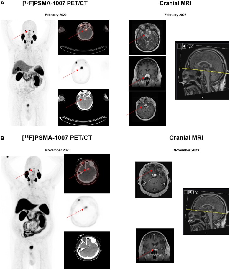
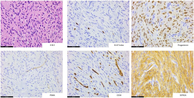
Prostate adenocarcinoma metastasis to brain has been reported to occur only up to 0.6% of patients and these are mostly diagnosed in autopsy series. In the setting of biochemical recurrence of prostate cancer, a suspected PSMA-avid (prostate-specific membrane antigen) lesion in the brain is still strongly suggestive of an intracranial metastasis of prostate cancer. This needs, however, a thoroughly recurrency work-up due to other potentially PSMA-avid cranial lesions, as PSMA initially was developed for the imaging of primary CNS tumours. We report of a challenging clinical case of a 71-year-old-patient with a strongly PSMA-avid lesion at the skull base. Given the medical history of a meningioma at the skull base, the further diagnostic work-up with MRI could still not rule out a malignancy, so that the patient needed to undergo a surgical excision of the tumour mass. The histological and immunohistochemical examinations revealed a relapsed CNS WHO grade 1 meningioma. From the aspect of molecular imaging and critical analysis of regular clinical care in a third-level university hospital, we consider this result very intriguing. Hence, we analyse the decision-making process and clinical course of this case in the light of molecular imaging findings.


 分享
分享
 求助内容:
求助内容: 应助结果提醒方式:
 扫码关注我们
