P. Slepička , N. Slepičková Kasálková , J. Musílková , L. Bačáková , B. Frýdlová , P. Sajdl , Z. Kolská , E. Rebollar , V. Švorčík
{"title":"PLLA honeycombs activated by plasma and high-energy excimer laser for stem cell support","authors":"P. Slepička , N. Slepičková Kasálková , J. Musílková , L. Bačáková , B. Frýdlová , P. Sajdl , Z. Kolská , E. Rebollar , V. Švorčík","doi":"10.1016/j.apsadv.2024.100662","DOIUrl":null,"url":null,"abstract":"<div><div>In this study, we constructed and activated honeycomb structures on perfluorinated substrates subjected to KrF laser treatment (wavelength 248 nm). We selected the biopolymer poly L-lactic acid (PLLA) as the honeycomb material, which was dissolved in a mixture of chloroform/methanol. A micropattern of a plasma-treated perfluorethyleneperopylene (FEP) substrate was prepared by improved phase separation during dip-coating. The PLLA micropattern was subsequently treated with an excimer laser with a laser fluence of 10 mJ.cm<sup>-2</sup> and with a different number of laser pulses. Alternatively, plasma exposure can be used as a secondary treatment. The surface morphologies of the pristine and laser-treated PLLA patterns were studied using atomic force and scanning electron microscopy. The surface chemistry was analyzed using energy-dispersive spectroscopy and X-ray photoelectron spectroscopy. In addition, the metabolic activity of adipose stem cells was evaluated using the MTS test, and cell numbers in selected samples were determined. The morphology of cells growing in the honeycomb-like pattern was studied in detail using fluorescence microscopy. In all, we used a combination of honeycomb pattern (HCP) laser treatment and plasma treatment to construct an optimal scaffold for adipose stem cell culture.</div></div>","PeriodicalId":34303,"journal":{"name":"Applied Surface Science Advances","volume":"25 ","pages":"Article 100662"},"PeriodicalIF":8.7000,"publicationDate":"2024-11-23","publicationTypes":"Journal Article","fieldsOfStudy":null,"isOpenAccess":false,"openAccessPdf":"","citationCount":"0","resultStr":null,"platform":"Semanticscholar","paperid":null,"PeriodicalName":"Applied Surface Science Advances","FirstCategoryId":"1085","ListUrlMain":"https://www.sciencedirect.com/science/article/pii/S2666523924000904","RegionNum":0,"RegionCategory":null,"ArticlePicture":[],"TitleCN":null,"AbstractTextCN":null,"PMCID":null,"EPubDate":"","PubModel":"","JCR":"Q1","JCRName":"CHEMISTRY, PHYSICAL","Score":null,"Total":0}
引用次数: 0
Abstract
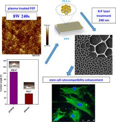
In this study, we constructed and activated honeycomb structures on perfluorinated substrates subjected to KrF laser treatment (wavelength 248 nm). We selected the biopolymer poly L-lactic acid (PLLA) as the honeycomb material, which was dissolved in a mixture of chloroform/methanol. A micropattern of a plasma-treated perfluorethyleneperopylene (FEP) substrate was prepared by improved phase separation during dip-coating. The PLLA micropattern was subsequently treated with an excimer laser with a laser fluence of 10 mJ.cm-2 and with a different number of laser pulses. Alternatively, plasma exposure can be used as a secondary treatment. The surface morphologies of the pristine and laser-treated PLLA patterns were studied using atomic force and scanning electron microscopy. The surface chemistry was analyzed using energy-dispersive spectroscopy and X-ray photoelectron spectroscopy. In addition, the metabolic activity of adipose stem cells was evaluated using the MTS test, and cell numbers in selected samples were determined. The morphology of cells growing in the honeycomb-like pattern was studied in detail using fluorescence microscopy. In all, we used a combination of honeycomb pattern (HCP) laser treatment and plasma treatment to construct an optimal scaffold for adipose stem cell culture.


 分享
分享
 求助内容:
求助内容: 应助结果提醒方式:
 扫码关注我们
