Comparative Effects of Crocin and Losartan on RAGE, TGF-β, TNF-α Gene Expression and Histopathological Changes of the Liver Tissue in Rats With Diabetes
{"title":"Comparative Effects of Crocin and Losartan on RAGE, TGF-β, TNF-α Gene Expression and Histopathological Changes of the Liver Tissue in Rats With Diabetes","authors":"Shahnaz Rajabi, Yaser Mohammadi, Hamid Kabiri-rad, Mahdiyeh Rajabi-moghaddam, Azam Rezaei Farimani","doi":"10.1002/edm2.70016","DOIUrl":null,"url":null,"abstract":"<div>\n \n \n <section>\n \n <h3> Background and Objectives</h3>\n \n <p>AGEs, via RAGE, increase the development of hyperglycemia-induced liver damage, and blocking this axis is associated with a reduction in liver disease progression. The goal of this study was to determine how crocin and losartan influenced RAGE, TNF-α and TGF-β gene expression in diabetic rats, as well as histological changes in liver tissue.</p>\n </section>\n \n <section>\n \n <h3> Materials and Methods</h3>\n \n <p>Diabetes was induced in 40 male Wistar rats using Streptozotocin (50 mg/kg, IP). There were five groups of rats: diabetic and healthy groups, diabetic rats given crocin (50 mg/kg), losartan (25 mg/kg) and both (crocin + Los). Serum glucose, ALT and AST levels were measured 4 weeks later. qPCR was used to examine the TNF-α, TGF-β and RAGE gene expression in liver tissue.</p>\n </section>\n \n <section>\n \n <h3> Results</h3>\n \n <p>Crocin was found to be effective in lowering FBG in the diabetes group. The serum levels of ALT and AST decreased in all treated groups, but this decrease was significant in the crocin + Los group (<i>p</i> < 0.05). The relative expression of RAGE, TNF-α and TGF-β genes was significantly higher in the diabetes group compared to the healthy group. The expression of these genes decreased in groups treated with crocin and Losartan compared to the diabetes group. The highest reduction in RAGE and TGF-β gene expression was reported in those treated with crocin + Los. Histopathology results showed that the diabetes group had more bile ducts and necrosis than the healthy control group, which had no tissue changes. Hepatocyte degeneration, bile duct proliferation, inflammatory changes and hepatocyte necrosis were mild in the treated groups, but no hepatocyte necrosis was observed in the crocin + Los group.</p>\n </section>\n \n <section>\n \n <h3> Conclusion</h3>\n \n <p>Crocin may be a feasible therapeutic agent for treating diabetes and its symptoms when combined with pharmaceutical medications. Human research is still needed to reach clear conclusions.</p>\n </section>\n </div>","PeriodicalId":36522,"journal":{"name":"Endocrinology, Diabetes and Metabolism","volume":"8 1","pages":""},"PeriodicalIF":2.6000,"publicationDate":"2024-11-28","publicationTypes":"Journal Article","fieldsOfStudy":null,"isOpenAccess":false,"openAccessPdf":"https://onlinelibrary.wiley.com/doi/epdf/10.1002/edm2.70016","citationCount":"0","resultStr":null,"platform":"Semanticscholar","paperid":null,"PeriodicalName":"Endocrinology, Diabetes and Metabolism","FirstCategoryId":"1085","ListUrlMain":"https://onlinelibrary.wiley.com/doi/10.1002/edm2.70016","RegionNum":0,"RegionCategory":null,"ArticlePicture":[],"TitleCN":null,"AbstractTextCN":null,"PMCID":null,"EPubDate":"","PubModel":"","JCR":"Q3","JCRName":"ENDOCRINOLOGY & METABOLISM","Score":null,"Total":0}
引用次数: 0
Abstract
Background and Objectives
AGEs, via RAGE, increase the development of hyperglycemia-induced liver damage, and blocking this axis is associated with a reduction in liver disease progression. The goal of this study was to determine how crocin and losartan influenced RAGE, TNF-α and TGF-β gene expression in diabetic rats, as well as histological changes in liver tissue.
Materials and Methods
Diabetes was induced in 40 male Wistar rats using Streptozotocin (50 mg/kg, IP). There were five groups of rats: diabetic and healthy groups, diabetic rats given crocin (50 mg/kg), losartan (25 mg/kg) and both (crocin + Los). Serum glucose, ALT and AST levels were measured 4 weeks later. qPCR was used to examine the TNF-α, TGF-β and RAGE gene expression in liver tissue.
Results
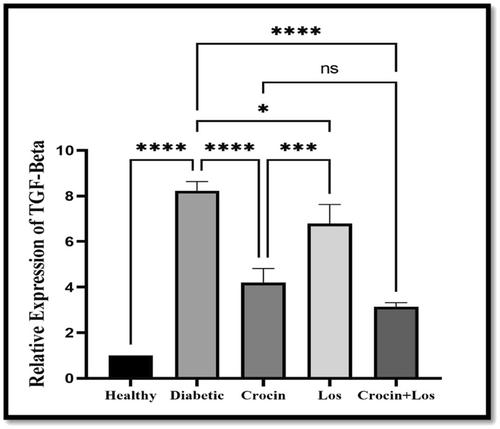
Crocin was found to be effective in lowering FBG in the diabetes group. The serum levels of ALT and AST decreased in all treated groups, but this decrease was significant in the crocin + Los group (p < 0.05). The relative expression of RAGE, TNF-α and TGF-β genes was significantly higher in the diabetes group compared to the healthy group. The expression of these genes decreased in groups treated with crocin and Losartan compared to the diabetes group. The highest reduction in RAGE and TGF-β gene expression was reported in those treated with crocin + Los. Histopathology results showed that the diabetes group had more bile ducts and necrosis than the healthy control group, which had no tissue changes. Hepatocyte degeneration, bile duct proliferation, inflammatory changes and hepatocyte necrosis were mild in the treated groups, but no hepatocyte necrosis was observed in the crocin + Los group.
Conclusion
Crocin may be a feasible therapeutic agent for treating diabetes and its symptoms when combined with pharmaceutical medications. Human research is still needed to reach clear conclusions.


 分享
分享
 求助内容:
求助内容: 应助结果提醒方式:
 扫码关注我们
