Cameron A Czerpak, Michael Saheb Kashaf, Brandon K Zimmerman, Rebecca Mirville, Nicolas C Gasquet, Harry A Quigley, Thao D Nguyen
{"title":"The Strain Response to Intraocular Pressure Increase in the Lamina Cribrosa of Control Subjects and Glaucoma Patients.","authors":"Cameron A Czerpak, Michael Saheb Kashaf, Brandon K Zimmerman, Rebecca Mirville, Nicolas C Gasquet, Harry A Quigley, Thao D Nguyen","doi":"10.1167/tvst.13.12.7","DOIUrl":null,"url":null,"abstract":"<p><strong>Purpose: </strong>The purpose of this study was to measure biomechanical strains in the lamina cribrosa (LC) of living human eyes undergoing intraocular pressure (IOP) increase.</p><p><strong>Methods: </strong>Healthy control subjects and patients with glaucoma underwent optical coherence tomographic (OCT) imaging of the LC before and after wearing of swim goggles that increased IOP (57 image pairs, 39 persons). Digital volume correlation was used to measure biomechanical strains in optic nerve head tissue and change in depth of the anterior border of the LC.</p><p><strong>Results: </strong>The mean IOP increase in both glaucoma and control eyes was 7.1 millimeters of mercury (mm Hg) after application of the goggles. Among glaucoma eyes, strains that were significant were: contractile Ezz (average = -0.33%, P = 0.0005), contractile Eθθ (average = -0.23%, P = 0.03), Emax (average = 0.83%, P < 0.0001), and Γmax (average = 0.95%, P < 0.0001), whereas the average anterior LC depth (ALD) decreased by 2.39 µm (anterior; P = 0.0002). In glaucoma eyes, shear strain Ezθ was greater with worse mean deviation (MD) and visual function index (P = 0.044 and P = 0.006, respectively, multivariate models). Strain compliance for Erθ, Ezθ, and Eθθ all increased with greater MD worsening prior to imaging (P = 0.04, P = 0.007, and P = 0.03).</p><p><strong>Conclusions: </strong>LC strains were measurable 20 minutes after IOP increase, producing axial compression and greater peripheral strain than centrally. Some strain compliances were greater with worse existing visual field loss or with more progressive past field loss.</p><p><strong>Translational relevance: </strong>Biomechanical strains are related to measures of glaucoma damage, supporting the hypothesis that optic nerve head biomechanical responses represent a noninvasive biomarker for glaucoma.</p>","PeriodicalId":23322,"journal":{"name":"Translational Vision Science & Technology","volume":"13 12","pages":"7"},"PeriodicalIF":2.6000,"publicationDate":"2024-12-02","publicationTypes":"Journal Article","fieldsOfStudy":null,"isOpenAccess":false,"openAccessPdf":"https://www.ncbi.nlm.nih.gov/pmc/articles/PMC11627119/pdf/","citationCount":"0","resultStr":null,"platform":"Semanticscholar","paperid":null,"PeriodicalName":"Translational Vision Science & Technology","FirstCategoryId":"3","ListUrlMain":"https://doi.org/10.1167/tvst.13.12.7","RegionNum":3,"RegionCategory":"医学","ArticlePicture":[],"TitleCN":null,"AbstractTextCN":null,"PMCID":null,"EPubDate":"","PubModel":"","JCR":"Q2","JCRName":"OPHTHALMOLOGY","Score":null,"Total":0}
引用次数: 0
Abstract
Purpose: The purpose of this study was to measure biomechanical strains in the lamina cribrosa (LC) of living human eyes undergoing intraocular pressure (IOP) increase.
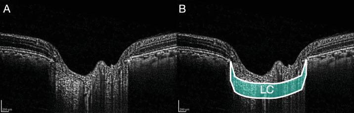
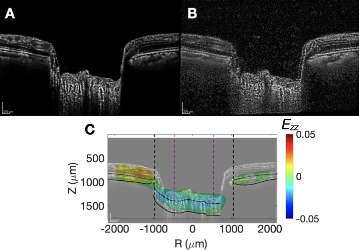
Methods: Healthy control subjects and patients with glaucoma underwent optical coherence tomographic (OCT) imaging of the LC before and after wearing of swim goggles that increased IOP (57 image pairs, 39 persons). Digital volume correlation was used to measure biomechanical strains in optic nerve head tissue and change in depth of the anterior border of the LC.
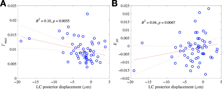
Results: The mean IOP increase in both glaucoma and control eyes was 7.1 millimeters of mercury (mm Hg) after application of the goggles. Among glaucoma eyes, strains that were significant were: contractile Ezz (average = -0.33%, P = 0.0005), contractile Eθθ (average = -0.23%, P = 0.03), Emax (average = 0.83%, P < 0.0001), and Γmax (average = 0.95%, P < 0.0001), whereas the average anterior LC depth (ALD) decreased by 2.39 µm (anterior; P = 0.0002). In glaucoma eyes, shear strain Ezθ was greater with worse mean deviation (MD) and visual function index (P = 0.044 and P = 0.006, respectively, multivariate models). Strain compliance for Erθ, Ezθ, and Eθθ all increased with greater MD worsening prior to imaging (P = 0.04, P = 0.007, and P = 0.03).
Conclusions: LC strains were measurable 20 minutes after IOP increase, producing axial compression and greater peripheral strain than centrally. Some strain compliances were greater with worse existing visual field loss or with more progressive past field loss.
Translational relevance: Biomechanical strains are related to measures of glaucoma damage, supporting the hypothesis that optic nerve head biomechanical responses represent a noninvasive biomarker for glaucoma.
期刊介绍:
Translational Vision Science & Technology (TVST), an official journal of the Association for Research in Vision and Ophthalmology (ARVO), an international organization whose purpose is to advance research worldwide into understanding the visual system and preventing, treating and curing its disorders, is an online, open access, peer-reviewed journal emphasizing multidisciplinary research that bridges the gap between basic research and clinical care. A highly qualified and diverse group of Associate Editors and Editorial Board Members is led by Editor-in-Chief Marco Zarbin, MD, PhD, FARVO.
The journal covers a broad spectrum of work, including but not limited to:
Applications of stem cell technology for regenerative medicine,
Development of new animal models of human diseases,
Tissue bioengineering,
Chemical engineering to improve virus-based gene delivery,
Nanotechnology for drug delivery,
Design and synthesis of artificial extracellular matrices,
Development of a true microsurgical operating environment,
Refining data analysis algorithms to improve in vivo imaging technology,
Results of Phase 1 clinical trials,
Reverse translational ("bedside to bench") research.
TVST seeks manuscripts from scientists and clinicians with diverse backgrounds ranging from basic chemistry to ophthalmic surgery that will advance or change the way we understand and/or treat vision-threatening diseases. TVST encourages the use of color, multimedia, hyperlinks, program code and other digital enhancements.


 分享
分享
 求助内容:
求助内容: 应助结果提醒方式:
 扫码关注我们
