Miguel A. Labrador-Espinosa, Jesús Silva-Rodriguez, Niels Okkels, Laura Muñoz-Delgado, Jacob Horsager, Sandra Castro-Labrador, Pablo Franco-Rosado, Ana María Castellano-Guerrero, Elena Iglesias-Camacho, Manuela San-Eufrasio, Daniel Macías-García, Silvia Jesús, Astrid Adarmes-Gómez, Elena Ojeda-Lepe, Fátima Carrillo, Juan Francisco Martín-Rodríguez, Florinda Roldan Lora, David García-Solís, Per Borghammer, Pablo Mir, Michel J. Grothe
{"title":"Cortical hypometabolism in Parkinson’s disease is linked to cholinergic basal forebrain atrophy","authors":"Miguel A. Labrador-Espinosa, Jesús Silva-Rodriguez, Niels Okkels, Laura Muñoz-Delgado, Jacob Horsager, Sandra Castro-Labrador, Pablo Franco-Rosado, Ana María Castellano-Guerrero, Elena Iglesias-Camacho, Manuela San-Eufrasio, Daniel Macías-García, Silvia Jesús, Astrid Adarmes-Gómez, Elena Ojeda-Lepe, Fátima Carrillo, Juan Francisco Martín-Rodríguez, Florinda Roldan Lora, David García-Solís, Per Borghammer, Pablo Mir, Michel J. Grothe","doi":"10.1038/s41380-024-02842-9","DOIUrl":null,"url":null,"abstract":"Cortical hypometabolism on FDG-PET is a well-established neuroimaging biomarker of cognitive impairment in Parkinson’s disease (PD), but its pathophysiologic origins are incompletely understood. Cholinergic basal forebrain (cBF) degeneration is a prominent pathological feature of PD-related cognitive impairment and may contribute to cortical hypometabolism through cholinergic denervation of cortical projection areas. Here, we investigated in-vivo associations between subregional cBF volumes on 3T-MRI, cortical hypometabolism on [18F]FDG-PET, and cognitive deficits in a cohort of 95 PD participants with varying degrees of cognitive impairment. We further assessed the spatial correspondence of the cortical pattern of cBF-associated hypometabolism with the pattern of cholinergic denervation in PD as assessed by [18F]FEOBV-PET imaging of presynaptic cholinergic terminal density in a second cohort. Lower volume of the cortically-projecting posterior cBF, but not of the anterior cBF, was significantly associated with extensive neocortical hypometabolism [p(FDR) < 0.05], which mediated the association between cBF atrophy and cognitive impairment (mediated proportion: 43%, p < 0.001). In combined models, posterior cBF atrophy explained more variance in cortical hypometabolism (R2 = 0.26, p < 0.001) than local atrophy in the cortical areas themselves (R2 = 0.16, p = 0.01). Topographic correspondence analysis with the [18F]FEOBV-PET pattern revealed that cortical areas showing most pronounced cBF-associated hypometabolism correspond to those showing most severe cholinergic denervation in PD (Spearman’s ρ = 0.57, p < 0.001). In conclusion, posterior cBF atrophy in PD is selectively associated with hypometabolism in denervated cortical target areas, which mediates the effect of cBF atrophy on cognitive impairment. These data provide first-time in-vivo evidence that cholinergic degeneration represents a principle pathological correlate of cortical hypometabolism underlying cognitive impairment in PD.","PeriodicalId":19008,"journal":{"name":"Molecular Psychiatry","volume":"30 6","pages":"2372-2380"},"PeriodicalIF":10.1000,"publicationDate":"2024-12-05","publicationTypes":"Journal Article","fieldsOfStudy":null,"isOpenAccess":false,"openAccessPdf":"","citationCount":"0","resultStr":null,"platform":"Semanticscholar","paperid":null,"PeriodicalName":"Molecular Psychiatry","FirstCategoryId":"3","ListUrlMain":"https://www.nature.com/articles/s41380-024-02842-9","RegionNum":1,"RegionCategory":"医学","ArticlePicture":[],"TitleCN":null,"AbstractTextCN":null,"PMCID":null,"EPubDate":"","PubModel":"","JCR":"Q1","JCRName":"BIOCHEMISTRY & MOLECULAR BIOLOGY","Score":null,"Total":0}
引用次数: 0
Abstract
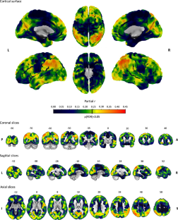
Cortical hypometabolism on FDG-PET is a well-established neuroimaging biomarker of cognitive impairment in Parkinson’s disease (PD), but its pathophysiologic origins are incompletely understood. Cholinergic basal forebrain (cBF) degeneration is a prominent pathological feature of PD-related cognitive impairment and may contribute to cortical hypometabolism through cholinergic denervation of cortical projection areas. Here, we investigated in-vivo associations between subregional cBF volumes on 3T-MRI, cortical hypometabolism on [18F]FDG-PET, and cognitive deficits in a cohort of 95 PD participants with varying degrees of cognitive impairment. We further assessed the spatial correspondence of the cortical pattern of cBF-associated hypometabolism with the pattern of cholinergic denervation in PD as assessed by [18F]FEOBV-PET imaging of presynaptic cholinergic terminal density in a second cohort. Lower volume of the cortically-projecting posterior cBF, but not of the anterior cBF, was significantly associated with extensive neocortical hypometabolism [p(FDR) < 0.05], which mediated the association between cBF atrophy and cognitive impairment (mediated proportion: 43%, p < 0.001). In combined models, posterior cBF atrophy explained more variance in cortical hypometabolism (R2 = 0.26, p < 0.001) than local atrophy in the cortical areas themselves (R2 = 0.16, p = 0.01). Topographic correspondence analysis with the [18F]FEOBV-PET pattern revealed that cortical areas showing most pronounced cBF-associated hypometabolism correspond to those showing most severe cholinergic denervation in PD (Spearman’s ρ = 0.57, p < 0.001). In conclusion, posterior cBF atrophy in PD is selectively associated with hypometabolism in denervated cortical target areas, which mediates the effect of cBF atrophy on cognitive impairment. These data provide first-time in-vivo evidence that cholinergic degeneration represents a principle pathological correlate of cortical hypometabolism underlying cognitive impairment in PD.
期刊介绍:
Molecular Psychiatry focuses on publishing research that aims to uncover the biological mechanisms behind psychiatric disorders and their treatment. The journal emphasizes studies that bridge pre-clinical and clinical research, covering cellular, molecular, integrative, clinical, imaging, and psychopharmacology levels.


 分享
分享
 求助内容:
求助内容: 应助结果提醒方式:
 扫码关注我们
