Jad Kassem, Ali Yildiz, Mamatha Gowda, Hunaina Shahab
{"title":"Dual left anterior descending artery: A case report.","authors":"Jad Kassem, Ali Yildiz, Mamatha Gowda, Hunaina Shahab","doi":"10.25259/JCIS_122_2024","DOIUrl":null,"url":null,"abstract":"<p><p>Dual left anterior descending (LAD) coronary artery is a rare anatomical variant with significant clinical implications. Recognizing this variant is crucial for accurate diagnosis and effective management, particularly in the context of revascularization strategies. We present a 71-year-old male with a history of dyspnea on exertion with baseline wall motion abnormality on a transthoracic stress echocardiography irreversible after exercise. Coronary computed tomography angiography revealed a dual LAD system: The left short LAD (LAD1) originating from the left main coronary artery and the right LAD (LAD2) arising separately from the right coronary cusp, distinct from the right coronary artery ostium. Having different origins and courses, both LADs supply the LAD territory. Our case is notable for two main reasons: The rarity of this particular type of dual LAD anatomy and the unique course of the LAD2, which, to our knowledge, has not been described in any previous case report. Although rare, dual LAD coronary artery should be considered in patients with atypical short LAD. Comprehensive imaging and a thorough understanding of coronary artery variants are essential for accurate diagnosis and effective management.</p>","PeriodicalId":15512,"journal":{"name":"Journal of Clinical Imaging Science","volume":"14 ","pages":"47"},"PeriodicalIF":1.3000,"publicationDate":"2024-12-03","publicationTypes":"Journal Article","fieldsOfStudy":null,"isOpenAccess":false,"openAccessPdf":"https://www.ncbi.nlm.nih.gov/pmc/articles/PMC11618701/pdf/","citationCount":"0","resultStr":null,"platform":"Semanticscholar","paperid":null,"PeriodicalName":"Journal of Clinical Imaging Science","FirstCategoryId":"1085","ListUrlMain":"https://doi.org/10.25259/JCIS_122_2024","RegionNum":0,"RegionCategory":null,"ArticlePicture":[],"TitleCN":null,"AbstractTextCN":null,"PMCID":null,"EPubDate":"2024/1/1 0:00:00","PubModel":"eCollection","JCR":"Q3","JCRName":"RADIOLOGY, NUCLEAR MEDICINE & MEDICAL IMAGING","Score":null,"Total":0}
引用次数: 0
Abstract
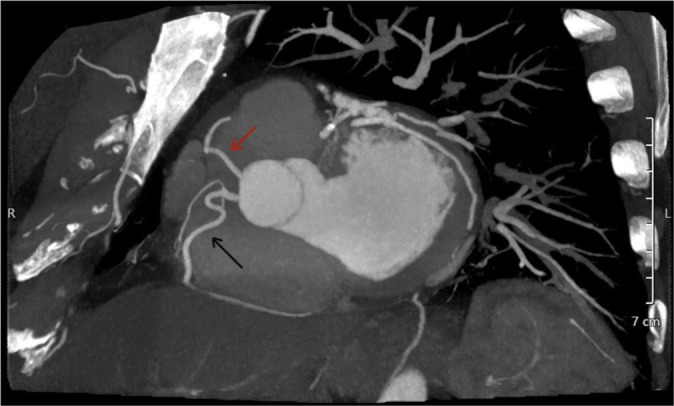
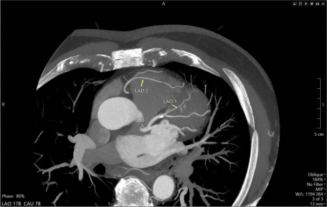
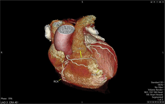
Dual left anterior descending (LAD) coronary artery is a rare anatomical variant with significant clinical implications. Recognizing this variant is crucial for accurate diagnosis and effective management, particularly in the context of revascularization strategies. We present a 71-year-old male with a history of dyspnea on exertion with baseline wall motion abnormality on a transthoracic stress echocardiography irreversible after exercise. Coronary computed tomography angiography revealed a dual LAD system: The left short LAD (LAD1) originating from the left main coronary artery and the right LAD (LAD2) arising separately from the right coronary cusp, distinct from the right coronary artery ostium. Having different origins and courses, both LADs supply the LAD territory. Our case is notable for two main reasons: The rarity of this particular type of dual LAD anatomy and the unique course of the LAD2, which, to our knowledge, has not been described in any previous case report. Although rare, dual LAD coronary artery should be considered in patients with atypical short LAD. Comprehensive imaging and a thorough understanding of coronary artery variants are essential for accurate diagnosis and effective management.
期刊介绍:
The Journal of Clinical Imaging Science (JCIS) is an open access peer-reviewed journal committed to publishing high-quality articles in the field of Imaging Science. The journal aims to present Imaging Science and relevant clinical information in an understandable and useful format. The journal is owned and published by the Scientific Scholar. Audience Our audience includes Radiologists, Researchers, Clinicians, medical professionals and students. Review process JCIS has a highly rigorous peer-review process that makes sure that manuscripts are scientifically accurate, relevant, novel and important. Authors disclose all conflicts, affiliations and financial associations such that the published content is not biased.


 分享
分享
 求助内容:
求助内容: 应助结果提醒方式:
 扫码关注我们
