Xuefei Guo, Haotian Li, Xiaoli Lu, Hao Liu, Kaicheng U, Chuangye Yan, Jianlin Lei, Jing Huang, Rui Zhou, Yigong Shi
{"title":"Structural basis of human γ-secretase inhibition by anticancer clinical compounds","authors":"Xuefei Guo, Haotian Li, Xiaoli Lu, Hao Liu, Kaicheng U, Chuangye Yan, Jianlin Lei, Jing Huang, Rui Zhou, Yigong Shi","doi":"10.1038/s41594-024-01439-8","DOIUrl":null,"url":null,"abstract":"Aberrant activation of Notch signaling, mediated by the Notch intracellular domain (NICD), is linked to certain types of cancer. The NICD is released through γ-secretase-mediated cleavage of the Notch receptor. Therefore, development of a γ-secretase inhibitor (GSI) represents an anticancer strategy. Here we report the cryo-electron microscopy structures of human γ-secretase bound individually to five clinically tested GSIs (RO4929097, crenigacestat, BMS906024, nirogacestat and MK-0752) at overall resolutions of 2.4–3.0 Å. Three of the five GSIs are in active anticancer clinical trials, while nirogacestat was recently approved. Each of these GSIs similarly occupies the substrate-binding site of presenilin 1 but shows characteristic differences in detailed recognition pattern. The size and shape of the binding pocket are induced by the bound GSI. Analysis of these structural features suggest strategies for modification of the GSI with improved inhibition potency. The cryo-electron microscopy structures of γ-secretase bound to five anticancer clinical compounds reveal characteristic differences in recognition and suggest strategies for improved drug design.","PeriodicalId":49141,"journal":{"name":"Nature Structural & Molecular Biology","volume":"32 4","pages":"719-728"},"PeriodicalIF":10.1000,"publicationDate":"2024-12-09","publicationTypes":"Journal Article","fieldsOfStudy":null,"isOpenAccess":false,"openAccessPdf":"","citationCount":"0","resultStr":null,"platform":"Semanticscholar","paperid":null,"PeriodicalName":"Nature Structural & Molecular Biology","FirstCategoryId":"99","ListUrlMain":"https://www.nature.com/articles/s41594-024-01439-8","RegionNum":1,"RegionCategory":"生物学","ArticlePicture":[],"TitleCN":null,"AbstractTextCN":null,"PMCID":null,"EPubDate":"","PubModel":"","JCR":"Q1","JCRName":"BIOCHEMISTRY & MOLECULAR BIOLOGY","Score":null,"Total":0}
引用次数: 0
Abstract
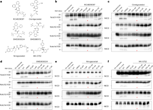
Aberrant activation of Notch signaling, mediated by the Notch intracellular domain (NICD), is linked to certain types of cancer. The NICD is released through γ-secretase-mediated cleavage of the Notch receptor. Therefore, development of a γ-secretase inhibitor (GSI) represents an anticancer strategy. Here we report the cryo-electron microscopy structures of human γ-secretase bound individually to five clinically tested GSIs (RO4929097, crenigacestat, BMS906024, nirogacestat and MK-0752) at overall resolutions of 2.4–3.0 Å. Three of the five GSIs are in active anticancer clinical trials, while nirogacestat was recently approved. Each of these GSIs similarly occupies the substrate-binding site of presenilin 1 but shows characteristic differences in detailed recognition pattern. The size and shape of the binding pocket are induced by the bound GSI. Analysis of these structural features suggest strategies for modification of the GSI with improved inhibition potency. The cryo-electron microscopy structures of γ-secretase bound to five anticancer clinical compounds reveal characteristic differences in recognition and suggest strategies for improved drug design.
期刊介绍:
Nature Structural & Molecular Biology is a comprehensive platform that combines structural and molecular research. Our journal focuses on exploring the functional and mechanistic aspects of biological processes, emphasizing how molecular components collaborate to achieve a particular function. While structural data can shed light on these insights, our publication does not require them as a prerequisite.


 分享
分享
 求助内容:
求助内容: 应助结果提醒方式:
 扫码关注我们
