Hebertt Gonzaga Dos Santos Chaves, Barbara Figueiredo, Caroline Andrade Maia, Alexandre Henrique Dos Reis-Prado, Maísa Mota Antunes, Ricardo Alves de Mesquita, Warley Luciano Fonseca Tavares, Gustavo Batista Menezes, Ivana Márcia Alves Diniz, Murilo Camuri Crovace, Gleide Fernandes de Avelar, Francine Benetti
{"title":"Tissue response and expression of interleukins (IL)-1ß, IL-6, IL-10 after pulp capping with bioglasses in mice.","authors":"Hebertt Gonzaga Dos Santos Chaves, Barbara Figueiredo, Caroline Andrade Maia, Alexandre Henrique Dos Reis-Prado, Maísa Mota Antunes, Ricardo Alves de Mesquita, Warley Luciano Fonseca Tavares, Gustavo Batista Menezes, Ivana Márcia Alves Diniz, Murilo Camuri Crovace, Gleide Fernandes de Avelar, Francine Benetti","doi":"10.1590/1807-3107bor-2024.vol38.0096","DOIUrl":null,"url":null,"abstract":"<p><p>This study aimed to evaluate the pulp response to F18 and cobalt-doped F18 bioglass (F18Co) in comparison with calcium hydroxide (CH) after pulp capping. The maxillary first molars of 48 rats were divided into F18, F18Co, CH, and control (no intervention) groups. The pulp was exposed, the materials were placed, and the teeth were capped. After 7 and 15 days, the animals were euthanized for pulp evaluation and interleukin (IL) expression determination. Statistical analysis was carried out using the SigmaPlot® program (Systat Software Inc., for Windows, version 12.0). The data obtained in the analyses were subjected to the non-parametric Kruskal-Wallis test, followed by Dunn's test. For all tests, statistical significance was set at p < 0.05. The CH group exhibited mild to moderate inflammation, whereas the bioglass groups displayed moderate to severe inflammation, indicating a notable difference between the control and bioglass groups. At 7 days, both the CH and most of the bioglass specimens showed moderate disorganization. On day 15, CH displayed mildto-moderate disorganization, whereas F18 and F18Co exhibited significantly more moderate-to-severe disorganization. There were no significant differences in IL-6 and IL-10 expressions between groups at 7 days, but a noteworthy increase in IL-1β was observed in both CH and F18. After 15 days, there was a greater expression of IL-6 and IL-1β in the bioglass groups. No significant IL-10 expression was observed. Bioglass performed less effectively than CH when in direct contact with the pulp tissue.</p>","PeriodicalId":9240,"journal":{"name":"Brazilian oral research","volume":"38 ","pages":"e096"},"PeriodicalIF":1.3000,"publicationDate":"2024-12-09","publicationTypes":"Journal Article","fieldsOfStudy":null,"isOpenAccess":false,"openAccessPdf":"https://www.ncbi.nlm.nih.gov/pmc/articles/PMC11654873/pdf/","citationCount":"0","resultStr":null,"platform":"Semanticscholar","paperid":null,"PeriodicalName":"Brazilian oral research","FirstCategoryId":"3","ListUrlMain":"https://doi.org/10.1590/1807-3107bor-2024.vol38.0096","RegionNum":4,"RegionCategory":"医学","ArticlePicture":[],"TitleCN":null,"AbstractTextCN":null,"PMCID":null,"EPubDate":"2024/1/1 0:00:00","PubModel":"eCollection","JCR":"Q3","JCRName":"DENTISTRY, ORAL SURGERY & MEDICINE","Score":null,"Total":0}
引用次数: 0
Abstract
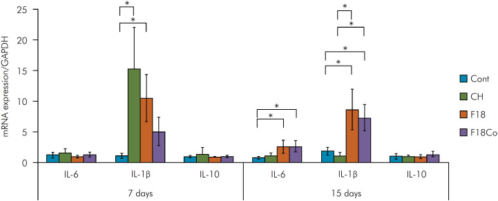
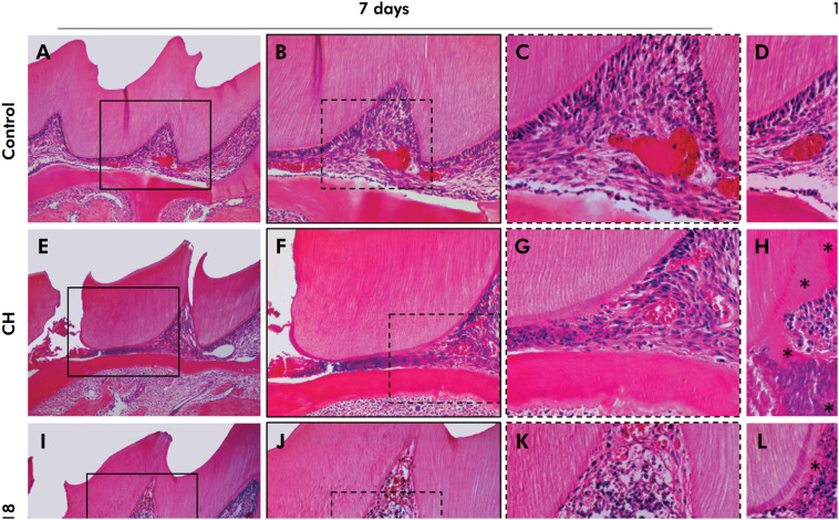
This study aimed to evaluate the pulp response to F18 and cobalt-doped F18 bioglass (F18Co) in comparison with calcium hydroxide (CH) after pulp capping. The maxillary first molars of 48 rats were divided into F18, F18Co, CH, and control (no intervention) groups. The pulp was exposed, the materials were placed, and the teeth were capped. After 7 and 15 days, the animals were euthanized for pulp evaluation and interleukin (IL) expression determination. Statistical analysis was carried out using the SigmaPlot® program (Systat Software Inc., for Windows, version 12.0). The data obtained in the analyses were subjected to the non-parametric Kruskal-Wallis test, followed by Dunn's test. For all tests, statistical significance was set at p < 0.05. The CH group exhibited mild to moderate inflammation, whereas the bioglass groups displayed moderate to severe inflammation, indicating a notable difference between the control and bioglass groups. At 7 days, both the CH and most of the bioglass specimens showed moderate disorganization. On day 15, CH displayed mildto-moderate disorganization, whereas F18 and F18Co exhibited significantly more moderate-to-severe disorganization. There were no significant differences in IL-6 and IL-10 expressions between groups at 7 days, but a noteworthy increase in IL-1β was observed in both CH and F18. After 15 days, there was a greater expression of IL-6 and IL-1β in the bioglass groups. No significant IL-10 expression was observed. Bioglass performed less effectively than CH when in direct contact with the pulp tissue.
本研究旨在评价牙髓封盖后对F18和钴掺杂F18生物玻璃(F18Co)的反应,并与氢氧化钙(CH)进行比较。48只大鼠上颌第一磨牙分为F18组、F18Co组、CH组和对照组(不干预组)。露出牙髓,放置材料,盖上牙齿。第7天和第15天处死动物,进行牙髓评估和白细胞介素(IL)表达测定。使用SigmaPlot®程序(Systat Software Inc., for Windows, version 12.0)进行统计分析。在分析中获得的数据进行非参数Kruskal-Wallis检验,然后进行Dunn检验。所有检验均以p < 0.05为差异有统计学意义。CH组表现为轻度至中度炎症,而生物玻璃组表现为中度至重度炎症,对照组与生物玻璃组之间存在显著差异。在第7天,CH和大部分生物玻璃标本都出现了中度的破坏。在第15天,CH表现出轻至中度的紊乱,而F18和F18Co表现出明显的中至重度紊乱。第7天各组间IL-6和IL-10的表达无显著差异,但CH和F18中IL-1β的表达均显著升高。15 d后,生物玻璃组IL-6和IL-1β表达增加。IL-10未见明显表达。当与牙髓组织直接接触时,生物玻璃的效果不如CH。


 分享
分享
 求助内容:
求助内容: 应助结果提醒方式:
 扫码关注我们
