{"title":"Diagnostic Value of Hepatic Mast Cell Concentration (MCC) in NAFLD and NASH Severity and Fibrosis Grade.","authors":"Mahshid Panahi, Nasser Rakhshani, Alireza Sarhaddi, Monavvar Afzalaghaee, Hamid Rezvani, Nikoo Emtiazi, Farkhonde Sarhaddi","doi":"10.30699/IJP.2024.2016320.3216","DOIUrl":null,"url":null,"abstract":"<p><strong>Background & objective: </strong>Mast cells play a role in the immune responses to fatty liver disease. The present study aimed to investigate the diagnostic value of hepatic mast cell concentration (MCC) in NAFLD and NASH severity and fibrosis grade.</p><p><strong>Methods: </strong>The present cross-sectional unremarkable hepatic histology, NAFLD, or NASH cases were enrolled. Demographic variables, BMI, hepatic stiffness assessed using fibroscan, portal inflammation, hepatic disease grade assessed using the NAFLD Activity Score (NAS), and hepatic fibrosis severity assessed using the NASH fibrosis stage, hepatic necrosis severity, and hepatic steatosis severity of the patients were collected. The hepatic specimens underwent immunohistochemical (IHC) staining.</p><p><strong>Results: </strong>Of a total of 92 patients with a mean age of 38.7±13.3 years, 56 (60.9%) were males. There were significant relationships between the NAS score of the patients and hepatic steatosis. Moreover, the NASH fibrosis stage had significant relationships with the variables of hepatic necrosis, steatosis, and stiffness. There were significant positive correlations between the mast cell concentration (MCC) in all zones of the hepatic tissue (zone 1, zone 2, zone 3, portal area, and total) and the variables of age, BMI, and hepatic necrosis, steatosis, and stiffness. The patients with a higher NASH fibrosis stage showed a significantly higher MCC in all zones of the hepatic tissue.</p><p><strong>Conclusion: </strong>Hepatic mast cell number may have a significant impact on the grade and fibrosis in NAFLD. However, it is recommended to perform further studies with larger sample sizes on patients with various etiologies for hepatic injury to confirm the present study results.</p>","PeriodicalId":38900,"journal":{"name":"Iranian Journal of Pathology","volume":"19 3","pages":"291-299"},"PeriodicalIF":0.0000,"publicationDate":"2024-01-01","publicationTypes":"Journal Article","fieldsOfStudy":null,"isOpenAccess":false,"openAccessPdf":"https://www.ncbi.nlm.nih.gov/pmc/articles/PMC11646207/pdf/","citationCount":"0","resultStr":null,"platform":"Semanticscholar","paperid":null,"PeriodicalName":"Iranian Journal of Pathology","FirstCategoryId":"1085","ListUrlMain":"https://doi.org/10.30699/IJP.2024.2016320.3216","RegionNum":0,"RegionCategory":null,"ArticlePicture":[],"TitleCN":null,"AbstractTextCN":null,"PMCID":null,"EPubDate":"2024/4/29 0:00:00","PubModel":"Epub","JCR":"Q3","JCRName":"Medicine","Score":null,"Total":0}
引用次数: 0
Abstract
Background & objective: Mast cells play a role in the immune responses to fatty liver disease. The present study aimed to investigate the diagnostic value of hepatic mast cell concentration (MCC) in NAFLD and NASH severity and fibrosis grade.
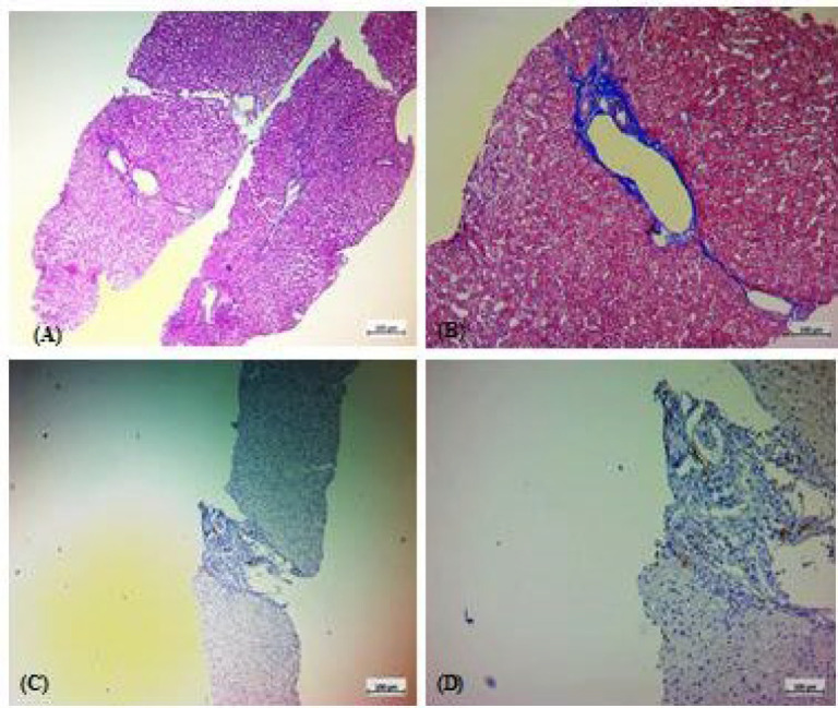
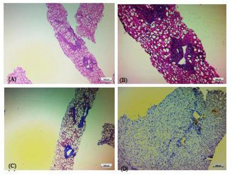
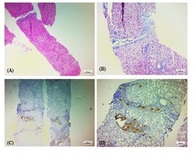
Methods: The present cross-sectional unremarkable hepatic histology, NAFLD, or NASH cases were enrolled. Demographic variables, BMI, hepatic stiffness assessed using fibroscan, portal inflammation, hepatic disease grade assessed using the NAFLD Activity Score (NAS), and hepatic fibrosis severity assessed using the NASH fibrosis stage, hepatic necrosis severity, and hepatic steatosis severity of the patients were collected. The hepatic specimens underwent immunohistochemical (IHC) staining.
Results: Of a total of 92 patients with a mean age of 38.7±13.3 years, 56 (60.9%) were males. There were significant relationships between the NAS score of the patients and hepatic steatosis. Moreover, the NASH fibrosis stage had significant relationships with the variables of hepatic necrosis, steatosis, and stiffness. There were significant positive correlations between the mast cell concentration (MCC) in all zones of the hepatic tissue (zone 1, zone 2, zone 3, portal area, and total) and the variables of age, BMI, and hepatic necrosis, steatosis, and stiffness. The patients with a higher NASH fibrosis stage showed a significantly higher MCC in all zones of the hepatic tissue.
Conclusion: Hepatic mast cell number may have a significant impact on the grade and fibrosis in NAFLD. However, it is recommended to perform further studies with larger sample sizes on patients with various etiologies for hepatic injury to confirm the present study results.


 分享
分享
 求助内容:
求助内容: 应助结果提醒方式:
 扫码关注我们
