Taha Yassine Ayadi , Amel Changuel , Hager Behi , Nabil Haloui , Karima Tlili , Med Bachir Khalifa
{"title":"Ectopic pancreatic tissue in the gallbladder: A rare incidental finding in a cholecystectomy specimen – A case report","authors":"Taha Yassine Ayadi , Amel Changuel , Hager Behi , Nabil Haloui , Karima Tlili , Med Bachir Khalifa","doi":"10.1016/j.ijscr.2024.110741","DOIUrl":null,"url":null,"abstract":"<div><h3>Introduction</h3><div>Ectopic pancreatic tissue (EPT) is a rare congenital anomaly characterized by the presence of pancreatic tissue in an abnormal location, separate from the pancreas, without any anatomical or vascular connection to it. This anomaly is often an incidental finding during operation or autopsy. This peculiarity poses clinical and radiological challenges for surgeons, particularly during laparoscopic or open procedures.</div></div><div><h3>Case report</h3><div>This article presents a compelling case of EPT, discovered incidentally during a planned laparoscopic cholecystectomy. The patient, a 50-year-old male with a history of biliary colic, underwent a meticulous laparoscopy exploration revealing an undistended gallbladder with an unexpected yellowish tissue fragment resembling pancreatic parenchyma.</div></div><div><h3>Clinical discussion</h3><div>EPT has an incidence ranging from 0.55 % to 13.7 % in autopsy studies. While the origins of EPT remain unclear, theories regarding the embryonic separation of pancreatic tissue offer insights into its origins and displacement from the original site. Macroscopically, EPT may present as polypoid lesions or yellow nodules and is typically asymptomatic. The various attachment locations and potential manifestations in other intra-abdominal sites add complexity to its diagnosis. Imaging techniques are often ineffective, making histopathological examination essential for diagnosis.</div></div><div><h3>Conclusion</h3><div>Diagnosing EPT in the gallbladder before and during surgery often presents significant challenges. Pathologists should be aware of this rare incidental finding, as it can mimic a tumor and lead to an overdiagnosis of malignancy. Only a precise histopathologic examination can provide a definite diagnosis and distinguish it from malignancies. Laparoscopic cholecystectomy is sufficing treatment.</div></div>","PeriodicalId":48113,"journal":{"name":"International Journal of Surgery Case Reports","volume":"126 ","pages":"Article 110741"},"PeriodicalIF":0.7000,"publicationDate":"2025-01-01","publicationTypes":"Journal Article","fieldsOfStudy":null,"isOpenAccess":false,"openAccessPdf":"https://www.ncbi.nlm.nih.gov/pmc/articles/PMC11718347/pdf/","citationCount":"0","resultStr":null,"platform":"Semanticscholar","paperid":null,"PeriodicalName":"International Journal of Surgery Case Reports","FirstCategoryId":"1085","ListUrlMain":"https://www.sciencedirect.com/science/article/pii/S2210261224015220","RegionNum":0,"RegionCategory":null,"ArticlePicture":[],"TitleCN":null,"AbstractTextCN":null,"PMCID":null,"EPubDate":"","PubModel":"","JCR":"Q4","JCRName":"SURGERY","Score":null,"Total":0}
引用次数: 0
Abstract
Introduction
Ectopic pancreatic tissue (EPT) is a rare congenital anomaly characterized by the presence of pancreatic tissue in an abnormal location, separate from the pancreas, without any anatomical or vascular connection to it. This anomaly is often an incidental finding during operation or autopsy. This peculiarity poses clinical and radiological challenges for surgeons, particularly during laparoscopic or open procedures.
Case report
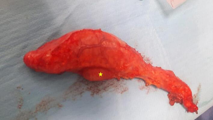
This article presents a compelling case of EPT, discovered incidentally during a planned laparoscopic cholecystectomy. The patient, a 50-year-old male with a history of biliary colic, underwent a meticulous laparoscopy exploration revealing an undistended gallbladder with an unexpected yellowish tissue fragment resembling pancreatic parenchyma.
Clinical discussion
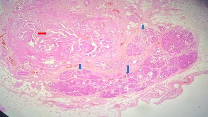
EPT has an incidence ranging from 0.55 % to 13.7 % in autopsy studies. While the origins of EPT remain unclear, theories regarding the embryonic separation of pancreatic tissue offer insights into its origins and displacement from the original site. Macroscopically, EPT may present as polypoid lesions or yellow nodules and is typically asymptomatic. The various attachment locations and potential manifestations in other intra-abdominal sites add complexity to its diagnosis. Imaging techniques are often ineffective, making histopathological examination essential for diagnosis.
Conclusion
Diagnosing EPT in the gallbladder before and during surgery often presents significant challenges. Pathologists should be aware of this rare incidental finding, as it can mimic a tumor and lead to an overdiagnosis of malignancy. Only a precise histopathologic examination can provide a definite diagnosis and distinguish it from malignancies. Laparoscopic cholecystectomy is sufficing treatment.


 分享
分享
 求助内容:
求助内容: 应助结果提醒方式:
 扫码关注我们
