Non-invasive removal of a misplaced and knotted guidewire during ultrasound-guided central venous catheter insertion in a hybrid operating room: a case report.
Mizuho Matsushita, Yoshikazu Yamaguchi, Honoka Yamashita, Chiyori Yamauchi, Hajime Hayami, Joseph D Tobias, Gaku Inagawa
{"title":"Non-invasive removal of a misplaced and knotted guidewire during ultrasound-guided central venous catheter insertion in a hybrid operating room: a case report.","authors":"Mizuho Matsushita, Yoshikazu Yamaguchi, Honoka Yamashita, Chiyori Yamauchi, Hajime Hayami, Joseph D Tobias, Gaku Inagawa","doi":"10.1186/s40981-024-00761-w","DOIUrl":null,"url":null,"abstract":"<p><strong>Background: </strong>The standard of care for placement of a central venous catheter (CVC) includes a real-time ultrasound (US)-guided technique. We describe a rare case in which the guidewire penetrated the posterior wall of the vessel, forming a knot, which precluded simple removal. This occurred despite the procedure being performed under real-time US guidance. The guidewire was eventually removed under fluoroscopic guidance in a hybrid operation room.</p><p><strong>Case presentation: </strong>An 89-year-old male underwent the placement of a CVC in the left internal jugular vein. During the US-guided procedure, the guidewire penetrated the posterior wall of the vessel and formed a knot, which impeded simple removal. This was confirmed by radiologic imaging. Using a short sheath and a push-pull technique, the radiologist was able to untangle the knot to allow for catheter removal. The guidewire was safely removed without vascular injury.</p><p><strong>Conclusions: </strong>A very rare complication of guidewire knotting was observed despite the use of US-guidance during needle and wire placement. The use of US, computed tomography, and fluoroscopy were beneficial for diagnosis, while the hybrid operating room provided the optimal environment for the removal procedure.</p>","PeriodicalId":14635,"journal":{"name":"JA Clinical Reports","volume":"10 1","pages":"78"},"PeriodicalIF":1.0000,"publicationDate":"2024-12-21","publicationTypes":"Journal Article","fieldsOfStudy":null,"isOpenAccess":false,"openAccessPdf":"https://www.ncbi.nlm.nih.gov/pmc/articles/PMC11663202/pdf/","citationCount":"0","resultStr":null,"platform":"Semanticscholar","paperid":null,"PeriodicalName":"JA Clinical Reports","FirstCategoryId":"1085","ListUrlMain":"https://doi.org/10.1186/s40981-024-00761-w","RegionNum":0,"RegionCategory":null,"ArticlePicture":[],"TitleCN":null,"AbstractTextCN":null,"PMCID":null,"EPubDate":"","PubModel":"","JCR":"Q3","JCRName":"ANESTHESIOLOGY","Score":null,"Total":0}
引用次数: 0
Abstract
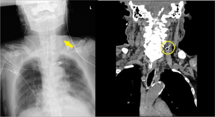
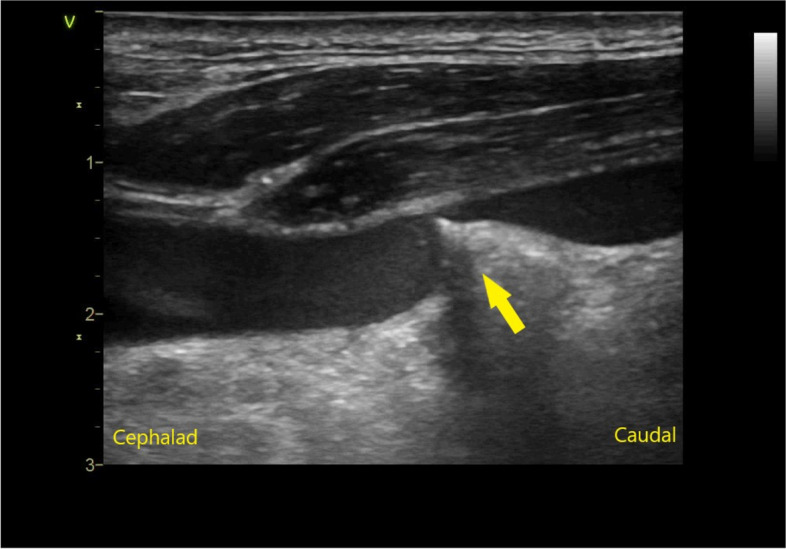
Background: The standard of care for placement of a central venous catheter (CVC) includes a real-time ultrasound (US)-guided technique. We describe a rare case in which the guidewire penetrated the posterior wall of the vessel, forming a knot, which precluded simple removal. This occurred despite the procedure being performed under real-time US guidance. The guidewire was eventually removed under fluoroscopic guidance in a hybrid operation room.
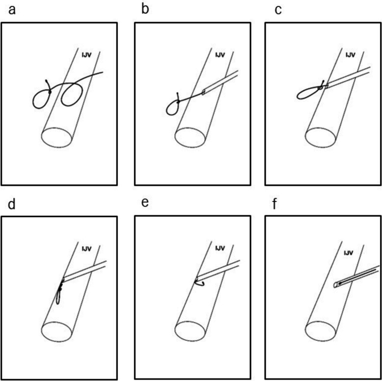
Case presentation: An 89-year-old male underwent the placement of a CVC in the left internal jugular vein. During the US-guided procedure, the guidewire penetrated the posterior wall of the vessel and formed a knot, which impeded simple removal. This was confirmed by radiologic imaging. Using a short sheath and a push-pull technique, the radiologist was able to untangle the knot to allow for catheter removal. The guidewire was safely removed without vascular injury.
Conclusions: A very rare complication of guidewire knotting was observed despite the use of US-guidance during needle and wire placement. The use of US, computed tomography, and fluoroscopy were beneficial for diagnosis, while the hybrid operating room provided the optimal environment for the removal procedure.
期刊介绍:
JA Clinical Reports is a companion journal to the Journal of Anesthesia (JA), the official journal of the Japanese Society of Anesthesiologists (JSA). This journal is an open access, peer-reviewed, online journal related to clinical anesthesia practices such as anesthesia management, pain management and intensive care. Case reports are very important articles from the viewpoint of education and the cultivation of scientific thinking in the field of anesthesia. However, submissions of anesthesia research and clinical reports from Japan are notably decreasing in major anesthesia journals. Therefore, the JSA has decided to launch a new journal, JA Clinical Reports, to encourage JSA members, particularly junior Japanese anesthesiologists, to publish papers in English language.


 分享
分享
 求助内容:
求助内容: 应助结果提醒方式:
 扫码关注我们
