{"title":"The structures of the peptide transporters SLC15A3 and SLC15A4 reveal the recognition mechanisms for substrate and TASL","authors":"Zhikuan Zhang, Shota Kasai, Kentaro Sakaniwa, Akiko Fujimura, Umeharu Ohto, Toshiyuki Shimizu","doi":"10.1016/j.str.2024.11.019","DOIUrl":null,"url":null,"abstract":"The solute carrier family 15 members 3 and 4 (SLC15A3 and SLC15A4) are closely related endolysosomal peptide transporters that transport free histidine and certain dipeptides from the lumen to cytosol. Besides, SLC15A4 also functions as a scaffold protein for the recruitment of the adapter TASL for interferon regulatory factor 5 (IRF5) activation downstream of innate immune TLR7-9 signaling. However, the molecular basis for the substrate recognition and TASL recruitment by these membrane proteins is not well understood. Here, we report the cryoelectron microscopy (cryo-EM) structure of apo SLC15A3 and structures of SLC15A4 in the absence or presence of the substrate, revealing the specific dipeptide recognition mechanism. Each SLC15A3 and SLC15A4 protomer adopts an outward-facing conformation. Furthermore, we also present the cryo-EM structure of a SLC15A4-TASL complex. The N terminal region of TASL forms a helical structure that inserts deeply into the inward-facing cavity of SLC15A4.","PeriodicalId":22168,"journal":{"name":"Structure","volume":"61 1","pages":""},"PeriodicalIF":4.3000,"publicationDate":"2024-12-23","publicationTypes":"Journal Article","fieldsOfStudy":null,"isOpenAccess":false,"openAccessPdf":"","citationCount":"0","resultStr":null,"platform":"Semanticscholar","paperid":null,"PeriodicalName":"Structure","FirstCategoryId":"99","ListUrlMain":"https://doi.org/10.1016/j.str.2024.11.019","RegionNum":2,"RegionCategory":"生物学","ArticlePicture":[],"TitleCN":null,"AbstractTextCN":null,"PMCID":null,"EPubDate":"","PubModel":"","JCR":"Q2","JCRName":"BIOCHEMISTRY & MOLECULAR BIOLOGY","Score":null,"Total":0}
引用次数: 0
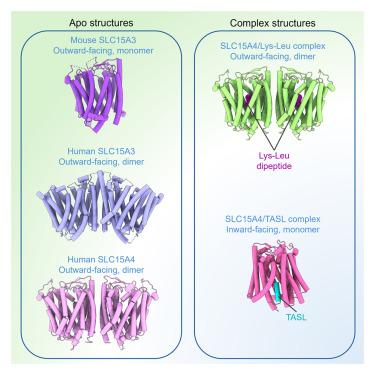
Abstract
The solute carrier family 15 members 3 and 4 (SLC15A3 and SLC15A4) are closely related endolysosomal peptide transporters that transport free histidine and certain dipeptides from the lumen to cytosol. Besides, SLC15A4 also functions as a scaffold protein for the recruitment of the adapter TASL for interferon regulatory factor 5 (IRF5) activation downstream of innate immune TLR7-9 signaling. However, the molecular basis for the substrate recognition and TASL recruitment by these membrane proteins is not well understood. Here, we report the cryoelectron microscopy (cryo-EM) structure of apo SLC15A3 and structures of SLC15A4 in the absence or presence of the substrate, revealing the specific dipeptide recognition mechanism. Each SLC15A3 and SLC15A4 protomer adopts an outward-facing conformation. Furthermore, we also present the cryo-EM structure of a SLC15A4-TASL complex. The N terminal region of TASL forms a helical structure that inserts deeply into the inward-facing cavity of SLC15A4.
期刊介绍:
Structure aims to publish papers of exceptional interest in the field of structural biology. The journal strives to be essential reading for structural biologists, as well as biologists and biochemists that are interested in macromolecular structure and function. Structure strongly encourages the submission of manuscripts that present structural and molecular insights into biological function and mechanism. Other reports that address fundamental questions in structural biology, such as structure-based examinations of protein evolution, folding, and/or design, will also be considered. We will consider the application of any method, experimental or computational, at high or low resolution, to conduct structural investigations, as long as the method is appropriate for the biological, functional, and mechanistic question(s) being addressed. Likewise, reports describing single-molecule analysis of biological mechanisms are welcome.
In general, the editors encourage submission of experimental structural studies that are enriched by an analysis of structure-activity relationships and will not consider studies that solely report structural information unless the structure or analysis is of exceptional and broad interest. Studies reporting only homology models, de novo models, or molecular dynamics simulations are also discouraged unless the models are informed by or validated by novel experimental data; rationalization of a large body of existing experimental evidence and making testable predictions based on a model or simulation is often not considered sufficient.


 分享
分享
 求助内容:
求助内容: 应助结果提醒方式:
 扫码关注我们
