{"title":"IK Channel Confers Fine-tuning of Rod Bipolar Cell Excitation and Synaptic Transmission in the Retina.","authors":"Yong Soo Park, Ki-Wug Sung, In-Beom Kim","doi":"10.1093/function/zqae054","DOIUrl":null,"url":null,"abstract":"<p><p>During retinal visual processing, rod bipolar cells (RBC) transfer scotopic signals from rods to AII amacrine cells as second-order neurons. Elucidation of the RBC's excitation/inhibition is essential for understanding the visual signal transmission. Excitation mechanisms via mGluR6 and voltage-gated Ca2+ channels in the RBCs and GABAergic inhibitory synaptic inputs have been studied in previous studies. However, its intrinsic inhibitory mechanisms like K+ and Cl- channels remain unclear. We focused on RBC's prominent K+ current, which exhibits voltage and Ca2+ dependence. We isolated and confirmed the expression of intermediate-conductance Ca2+-activated K+ channels (IK) in RBCs using the patch-clamp method with IK inhibitors (clotrimazole and TRAM34) and immunohistochemistry. The regulation of the IK channel primarily relies on Ca2+ influx via low-threshold Ca2+ channels during RBC's excitation. Additionally, IK mediates late repolarization and suppresses excessive oscillation of the membrane potential in the RBCs, enabling fast and transient synaptic transmission to AII amacrine cells. Our findings highlight the unique role of the IK channel in RBCs, suggesting that it plays a critical role in the scotopic pathway by fine-tuning RBC activity.</p>","PeriodicalId":73119,"journal":{"name":"Function (Oxford, England)","volume":" ","pages":""},"PeriodicalIF":3.8000,"publicationDate":"2025-02-12","publicationTypes":"Journal Article","fieldsOfStudy":null,"isOpenAccess":false,"openAccessPdf":"https://www.ncbi.nlm.nih.gov/pmc/articles/PMC11815585/pdf/","citationCount":"0","resultStr":null,"platform":"Semanticscholar","paperid":null,"PeriodicalName":"Function (Oxford, England)","FirstCategoryId":"1085","ListUrlMain":"https://doi.org/10.1093/function/zqae054","RegionNum":0,"RegionCategory":null,"ArticlePicture":[],"TitleCN":null,"AbstractTextCN":null,"PMCID":null,"EPubDate":"","PubModel":"","JCR":"Q2","JCRName":"CELL BIOLOGY","Score":null,"Total":0}
引用次数: 0
Abstract
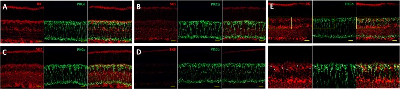
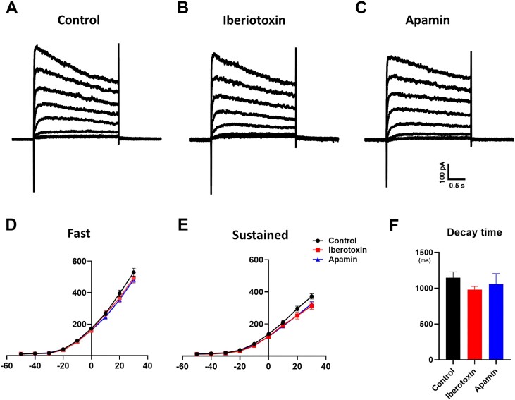
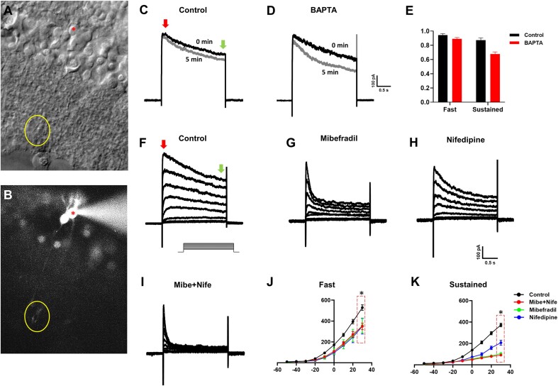
During retinal visual processing, rod bipolar cells (RBC) transfer scotopic signals from rods to AII amacrine cells as second-order neurons. Elucidation of the RBC's excitation/inhibition is essential for understanding the visual signal transmission. Excitation mechanisms via mGluR6 and voltage-gated Ca2+ channels in the RBCs and GABAergic inhibitory synaptic inputs have been studied in previous studies. However, its intrinsic inhibitory mechanisms like K+ and Cl- channels remain unclear. We focused on RBC's prominent K+ current, which exhibits voltage and Ca2+ dependence. We isolated and confirmed the expression of intermediate-conductance Ca2+-activated K+ channels (IK) in RBCs using the patch-clamp method with IK inhibitors (clotrimazole and TRAM34) and immunohistochemistry. The regulation of the IK channel primarily relies on Ca2+ influx via low-threshold Ca2+ channels during RBC's excitation. Additionally, IK mediates late repolarization and suppresses excessive oscillation of the membrane potential in the RBCs, enabling fast and transient synaptic transmission to AII amacrine cells. Our findings highlight the unique role of the IK channel in RBCs, suggesting that it plays a critical role in the scotopic pathway by fine-tuning RBC activity.


 分享
分享

 求助内容:
求助内容: 应助结果提醒方式:
 扫码关注我们
