{"title":"Anatomical Variations of the Vermiform Appendix.","authors":"Athanasios Sakellariadis, Foteini Sofou, Dimosthenis Chrysikos, Theodoros Sampsakos-Mariolis, Dimitrios Schizas, Theodoros Troupis, Dimitrios Filippou","doi":"10.5644/ama2006-124.461","DOIUrl":null,"url":null,"abstract":"<p><strong>Objective: </strong>The aim of the present work is to systematically review and present the existing literature on anatomical variations of the appendix.</p><p><strong>Methods: </strong>Detailed research was conducted in the PubMed medical database, using the terms \"Appendix\" AND \"Anatomical variations\", and 74 articles were initially revealed. After the application of the inclusion and exclusion criteria, all the non-related articles were excluded, and thus 40 articles were finally selected.</p><p><strong>Discussion: </strong>The data analysis suggests that the location and form of the appendix may significantly vary among individuals. Common anatomical variations concerning its location include retrocecal, pelvic, retro-ileal, pre-ileal, prececal and paracecal appendices. The first two variants are the most common, although there is a discrepancy regarding their exact incidence. Rarely, the appendix may be intracecal, intramural, subhepatic or located in the left abdomen; mismatches of the McBurney guide point with the base of the appendix are also recorded. Concerning the appendix's form, several variations in the length, diameter, shape and number of appendages (doubling, tripling) may be present.</p><p><strong>Conclusions: </strong>As evident from the presentation of the results, the vermiform appendix presents a wide variety and number of anatomical variations. The latter are of particular clinical importance and should be known to doctors - especially surgeons - to avoid complications in clinical practice.</p>","PeriodicalId":38313,"journal":{"name":"Acta medica academica","volume":" ","pages":"335-342"},"PeriodicalIF":0.0000,"publicationDate":"2024-12-01","publicationTypes":"Journal Article","fieldsOfStudy":null,"isOpenAccess":false,"openAccessPdf":"https://www.ncbi.nlm.nih.gov/pmc/articles/PMC11831545/pdf/","citationCount":"0","resultStr":null,"platform":"Semanticscholar","paperid":null,"PeriodicalName":"Acta medica academica","FirstCategoryId":"1085","ListUrlMain":"https://doi.org/10.5644/ama2006-124.461","RegionNum":0,"RegionCategory":null,"ArticlePicture":[],"TitleCN":null,"AbstractTextCN":null,"PMCID":null,"EPubDate":"","PubModel":"","JCR":"Q3","JCRName":"Medicine","Score":null,"Total":0}
引用次数: 0
Abstract
Objective: The aim of the present work is to systematically review and present the existing literature on anatomical variations of the appendix.
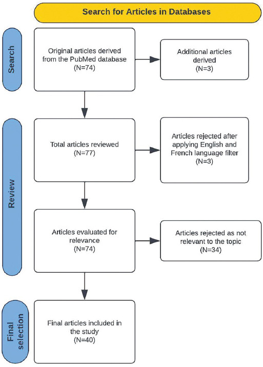
Methods: Detailed research was conducted in the PubMed medical database, using the terms "Appendix" AND "Anatomical variations", and 74 articles were initially revealed. After the application of the inclusion and exclusion criteria, all the non-related articles were excluded, and thus 40 articles were finally selected.
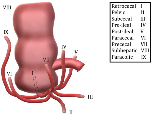
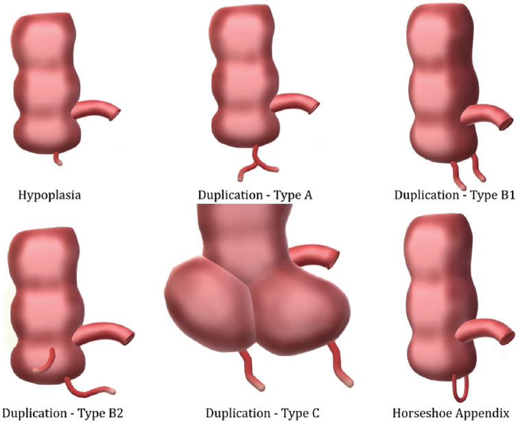
Discussion: The data analysis suggests that the location and form of the appendix may significantly vary among individuals. Common anatomical variations concerning its location include retrocecal, pelvic, retro-ileal, pre-ileal, prececal and paracecal appendices. The first two variants are the most common, although there is a discrepancy regarding their exact incidence. Rarely, the appendix may be intracecal, intramural, subhepatic or located in the left abdomen; mismatches of the McBurney guide point with the base of the appendix are also recorded. Concerning the appendix's form, several variations in the length, diameter, shape and number of appendages (doubling, tripling) may be present.
Conclusions: As evident from the presentation of the results, the vermiform appendix presents a wide variety and number of anatomical variations. The latter are of particular clinical importance and should be known to doctors - especially surgeons - to avoid complications in clinical practice.


 分享
分享
 求助内容:
求助内容: 应助结果提醒方式:
 扫码关注我们
