Sanghoon Kang , Jesus D. Penaloza Aponte , Omar Elashkar , Juan Francisco Morales , Nicholas Waddington , Damon G. Lamb , Huiwen Ju , Martha Campbell-Thompson , Sarah Kim
{"title":"Leveraging pre-trained machine learning models for islet quantification in type 1 diabetes","authors":"Sanghoon Kang , Jesus D. Penaloza Aponte , Omar Elashkar , Juan Francisco Morales , Nicholas Waddington , Damon G. Lamb , Huiwen Ju , Martha Campbell-Thompson , Sarah Kim","doi":"10.1016/j.jpi.2024.100406","DOIUrl":null,"url":null,"abstract":"<div><div>Human islets display a high degree of heterogeneity in terms of size, number, architecture, and endocrine cell-type compositions. An ever-increasing number of immunohistochemistry-stained whole slide images (WSIs) are available through the online pathology database of the Network for Pancreatic Organ donors with Diabetes (nPOD) program at the University of Florida (UF). We aimed to develop an enhanced machine learning-assisted WSI analysis workflow to utilize the nPOD resource for analysis of endocrine cell heterogeneity in the natural history of type 1 diabetes (T1D) in comparison to donors without diabetes. To maximize usability, the user-friendly open-source software QuPath was selected for the main interface. The WSI data were analyzed with two pre-trained machine learning models (i.e., Segment Anything Model (SAM) and QuPath's pixel classifier), using the UF high-performance-computing cluster, HiPerGator. SAM was used to define precise endocrine cell and cell grouping boundaries (with an average quality score of 0.91 per slide), and the artificial neural network-based pixel classifier was applied to segment areas of insulin- or glucagon-stained cytoplasmic regions within each endocrine cell. An additional script was developed to automatically count CD3+ cells inside and within 20 μm of each islet perimeter to quantify the number of islets with inflammation (i.e., CD3+ T-cell infiltration). Proof-of-concept analysis was performed to test the developed workflow in 12 subjects using 24 slides. This open-source machine learning-assisted workflow enables rapid and high throughput determinations of endocrine cells, whether as single cells or within groups, across hundreds of slides. It is expected that the use of this workflow will accelerate our understanding of endocrine cell and islet heterogeneity in the context of T1D endotypes and pathogenesis.</div></div>","PeriodicalId":37769,"journal":{"name":"Journal of Pathology Informatics","volume":"16 ","pages":"Article 100406"},"PeriodicalIF":0.0000,"publicationDate":"2025-01-01","publicationTypes":"Journal Article","fieldsOfStudy":null,"isOpenAccess":false,"openAccessPdf":"https://www.ncbi.nlm.nih.gov/pmc/articles/PMC11665367/pdf/","citationCount":"0","resultStr":null,"platform":"Semanticscholar","paperid":null,"PeriodicalName":"Journal of Pathology Informatics","FirstCategoryId":"1085","ListUrlMain":"https://www.sciencedirect.com/science/article/pii/S2153353924000452","RegionNum":0,"RegionCategory":null,"ArticlePicture":[],"TitleCN":null,"AbstractTextCN":null,"PMCID":null,"EPubDate":"","PubModel":"","JCR":"Q2","JCRName":"Medicine","Score":null,"Total":0}
引用次数: 0
Abstract
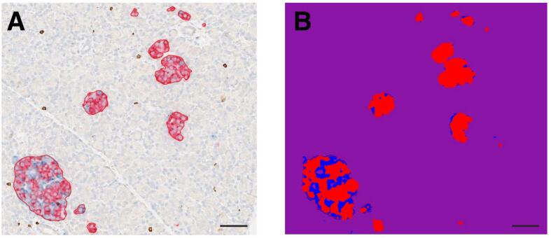
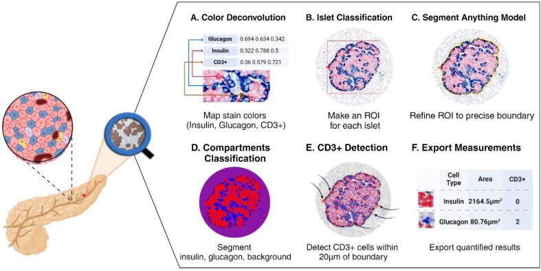
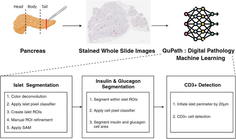
Human islets display a high degree of heterogeneity in terms of size, number, architecture, and endocrine cell-type compositions. An ever-increasing number of immunohistochemistry-stained whole slide images (WSIs) are available through the online pathology database of the Network for Pancreatic Organ donors with Diabetes (nPOD) program at the University of Florida (UF). We aimed to develop an enhanced machine learning-assisted WSI analysis workflow to utilize the nPOD resource for analysis of endocrine cell heterogeneity in the natural history of type 1 diabetes (T1D) in comparison to donors without diabetes. To maximize usability, the user-friendly open-source software QuPath was selected for the main interface. The WSI data were analyzed with two pre-trained machine learning models (i.e., Segment Anything Model (SAM) and QuPath's pixel classifier), using the UF high-performance-computing cluster, HiPerGator. SAM was used to define precise endocrine cell and cell grouping boundaries (with an average quality score of 0.91 per slide), and the artificial neural network-based pixel classifier was applied to segment areas of insulin- or glucagon-stained cytoplasmic regions within each endocrine cell. An additional script was developed to automatically count CD3+ cells inside and within 20 μm of each islet perimeter to quantify the number of islets with inflammation (i.e., CD3+ T-cell infiltration). Proof-of-concept analysis was performed to test the developed workflow in 12 subjects using 24 slides. This open-source machine learning-assisted workflow enables rapid and high throughput determinations of endocrine cells, whether as single cells or within groups, across hundreds of slides. It is expected that the use of this workflow will accelerate our understanding of endocrine cell and islet heterogeneity in the context of T1D endotypes and pathogenesis.
期刊介绍:
The Journal of Pathology Informatics (JPI) is an open access peer-reviewed journal dedicated to the advancement of pathology informatics. This is the official journal of the Association for Pathology Informatics (API). The journal aims to publish broadly about pathology informatics and freely disseminate all articles worldwide. This journal is of interest to pathologists, informaticians, academics, researchers, health IT specialists, information officers, IT staff, vendors, and anyone with an interest in informatics. We encourage submissions from anyone with an interest in the field of pathology informatics. We publish all types of papers related to pathology informatics including original research articles, technical notes, reviews, viewpoints, commentaries, editorials, symposia, meeting abstracts, book reviews, and correspondence to the editors. All submissions are subject to rigorous peer review by the well-regarded editorial board and by expert referees in appropriate specialties.


 分享
分享
 求助内容:
求助内容: 应助结果提醒方式:
 扫码关注我们
