Ali Fahd, Aya A Hussien, Mohamed T Ellabban, Zein A Shatat
{"title":"Validation of novel measurement protocols proposed for the standardized assessment of crestal bone levels: A cone-beam computed tomography study.","authors":"Ali Fahd, Aya A Hussien, Mohamed T Ellabban, Zein A Shatat","doi":"10.5624/isd.20240073","DOIUrl":null,"url":null,"abstract":"<p><strong>Purpose: </strong>This study was performed to introduce, evaluate, and compare various novel assessment protocols designed for straightforward, reliable, and reproducible measurement of alveolar bone levels. These protocols are intended for standardized periodontal assessment and follow-up, utilizing cone-beam computed tomography (CBCT) images and manipulation of Digital Imaging and Communications in Medicine (DICOM) viewer software.</p><p><strong>Materials and methods: </strong>Two experienced oral and maxillofacial radiologists developed 5 distinct radiographic measurement protocols. These techniques were established to assess the alveolar bone level of a periodontally affected upper central incisor using a method that is consistently repeatable across observers. Two additional assessors, blinded to the details of the study, independently applied the protocols to retrieved DICOM files that met the eligibility criteria. A scoring system with 3 subscores was created and used to compare the protocols.</p><p><strong>Results: </strong>Statistically excellent inter-observer reliability was observed for all protocols, other than protocol 1, which demonstrated moderate reliability. The average discrepancy between measurements taken by the 2 observers was 1.2 mm for protocol 1, 0.81 mm for protocol 2, and less than 0.5 mm for the remaining 3 protocols. All approaches except protocol 4 were straightforward to apply.</p><p><strong>Conclusion: </strong>This study introduces multiple reliable protocols for the evaluation of periodontal bone levels that ensure consistency across observers. Based on the findings, the double axial lines and incisocrestal distance protocols are recommended. These new assessment approaches, along with any future modifications, may be useful in periodontal assessment, dental implant follow-up, orthodontic evaluation, research, and artificial intelligence model generation.</p>","PeriodicalId":51714,"journal":{"name":"Imaging Science in Dentistry","volume":"54 4","pages":"354-361"},"PeriodicalIF":2.1000,"publicationDate":"2024-12-01","publicationTypes":"Journal Article","fieldsOfStudy":null,"isOpenAccess":false,"openAccessPdf":"https://www.ncbi.nlm.nih.gov/pmc/articles/PMC11685304/pdf/","citationCount":"0","resultStr":null,"platform":"Semanticscholar","paperid":null,"PeriodicalName":"Imaging Science in Dentistry","FirstCategoryId":"1085","ListUrlMain":"https://doi.org/10.5624/isd.20240073","RegionNum":0,"RegionCategory":null,"ArticlePicture":[],"TitleCN":null,"AbstractTextCN":null,"PMCID":null,"EPubDate":"2024/8/12 0:00:00","PubModel":"Epub","JCR":"Q3","JCRName":"DENTISTRY, ORAL SURGERY & MEDICINE","Score":null,"Total":0}
引用次数: 0
Abstract
Purpose: This study was performed to introduce, evaluate, and compare various novel assessment protocols designed for straightforward, reliable, and reproducible measurement of alveolar bone levels. These protocols are intended for standardized periodontal assessment and follow-up, utilizing cone-beam computed tomography (CBCT) images and manipulation of Digital Imaging and Communications in Medicine (DICOM) viewer software.
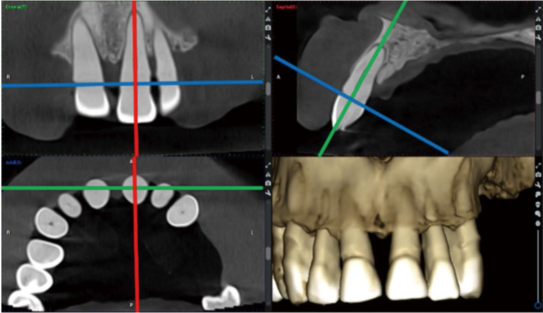
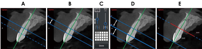
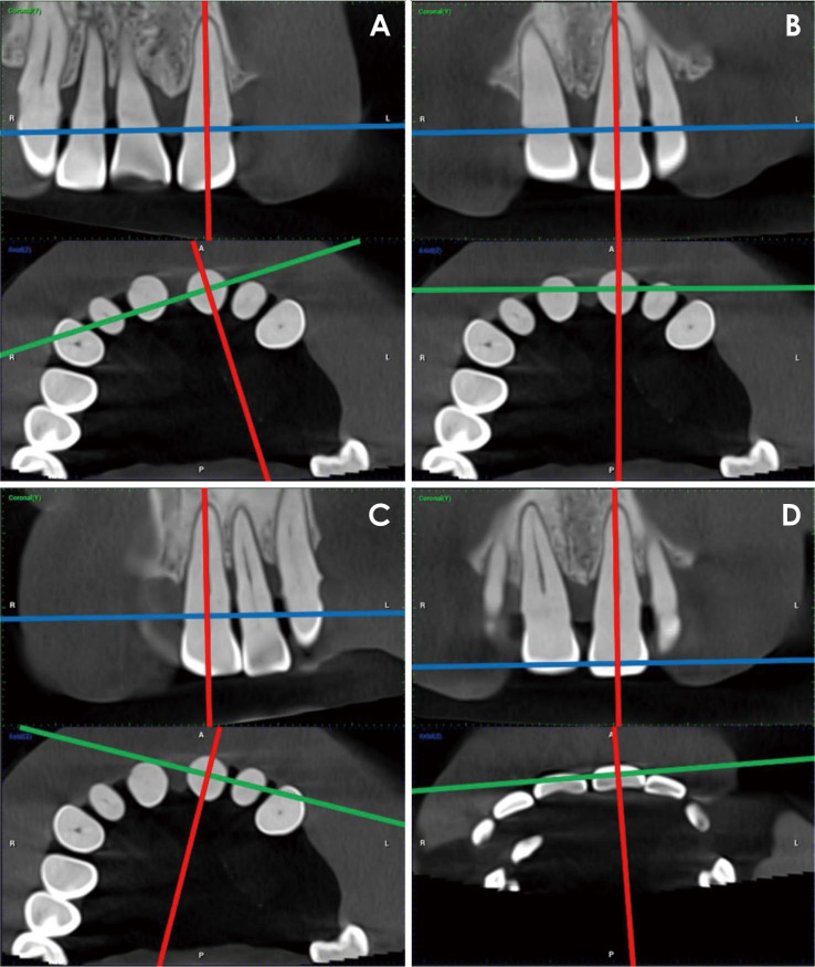
Materials and methods: Two experienced oral and maxillofacial radiologists developed 5 distinct radiographic measurement protocols. These techniques were established to assess the alveolar bone level of a periodontally affected upper central incisor using a method that is consistently repeatable across observers. Two additional assessors, blinded to the details of the study, independently applied the protocols to retrieved DICOM files that met the eligibility criteria. A scoring system with 3 subscores was created and used to compare the protocols.
Results: Statistically excellent inter-observer reliability was observed for all protocols, other than protocol 1, which demonstrated moderate reliability. The average discrepancy between measurements taken by the 2 observers was 1.2 mm for protocol 1, 0.81 mm for protocol 2, and less than 0.5 mm for the remaining 3 protocols. All approaches except protocol 4 were straightforward to apply.
Conclusion: This study introduces multiple reliable protocols for the evaluation of periodontal bone levels that ensure consistency across observers. Based on the findings, the double axial lines and incisocrestal distance protocols are recommended. These new assessment approaches, along with any future modifications, may be useful in periodontal assessment, dental implant follow-up, orthodontic evaluation, research, and artificial intelligence model generation.


 分享
分享
 求助内容:
求助内容: 应助结果提醒方式:
 扫码关注我们
