{"title":"Unusual presentation of esophageal tuberculosis: a case study.","authors":"Ming Xue, Yue-Can Zeng","doi":"10.1186/s12879-024-10418-9","DOIUrl":null,"url":null,"abstract":"<p><strong>Background: </strong>Esophageal ulcers can arise not only from malignant lesions but also from benign diseases, such as tuberculosis. These ulcers may mimic the radiological features of esophageal malignancy or tuberculosis on PET/CT, leading to diagnostic challenges.</p><p><strong>Case presentation: </strong>A 59-year-old woman was admitted to our hospital with a month-long history of progressive painful swallowing, fatigue, and loss of appetite. Whole-body 18 F-FDG PET/CT revealed a lesion in the mid-esophagus and swollen mediastinal lymph nodes with high FDG uptake, showing a maximum standardized uptake value (SUVmax) of 17.10 for the lymph nodes and 8.08 for the esophageal lesion. Esophageal cancer was initially suspected based on PET/CT findings. However, pathological examination of the esophageal lesion obtained via esophagoscopy showed only inflammation and granulation tissue, with no malignancy. A biopsy of the lymph nodes obtained through endoscopic ultrasonography revealed caseous necrosis but no atypical cells, and microbiological tests were positive for Mycobacterium tuberculosis. A final diagnosis of esophageal tuberculosis was made.</p><p><strong>Conclusions: </strong>Esophageal lesions can result from both malignant and benign conditions, including tuberculosis, and may mimic the radiological features of esophageal malignancy on PET/CT or other imaging studies. When esophageal lesions resemble malignancy, pseudotumoral esophagus and esophageal tuberculosis should be considered as differential diagnoses. Endoscopy, particularly endoscopic ultrasonography, is strongly recommended to accurately distinguish between benign and malignant esophageal lesions, helping to avoid unnecessary invasive treatments and reduce potential physical and psychological harm to patients.</p>","PeriodicalId":8981,"journal":{"name":"BMC Infectious Diseases","volume":"25 1","pages":"4"},"PeriodicalIF":3.2000,"publicationDate":"2025-01-02","publicationTypes":"Journal Article","fieldsOfStudy":null,"isOpenAccess":false,"openAccessPdf":"https://www.ncbi.nlm.nih.gov/pmc/articles/PMC11694470/pdf/","citationCount":"0","resultStr":null,"platform":"Semanticscholar","paperid":null,"PeriodicalName":"BMC Infectious Diseases","FirstCategoryId":"3","ListUrlMain":"https://doi.org/10.1186/s12879-024-10418-9","RegionNum":3,"RegionCategory":"医学","ArticlePicture":[],"TitleCN":null,"AbstractTextCN":null,"PMCID":null,"EPubDate":"","PubModel":"","JCR":"Q2","JCRName":"INFECTIOUS DISEASES","Score":null,"Total":0}
引用次数: 0
Abstract
Background: Esophageal ulcers can arise not only from malignant lesions but also from benign diseases, such as tuberculosis. These ulcers may mimic the radiological features of esophageal malignancy or tuberculosis on PET/CT, leading to diagnostic challenges.
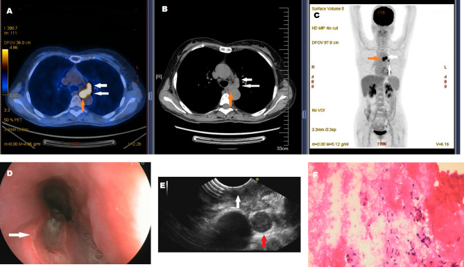
Case presentation: A 59-year-old woman was admitted to our hospital with a month-long history of progressive painful swallowing, fatigue, and loss of appetite. Whole-body 18 F-FDG PET/CT revealed a lesion in the mid-esophagus and swollen mediastinal lymph nodes with high FDG uptake, showing a maximum standardized uptake value (SUVmax) of 17.10 for the lymph nodes and 8.08 for the esophageal lesion. Esophageal cancer was initially suspected based on PET/CT findings. However, pathological examination of the esophageal lesion obtained via esophagoscopy showed only inflammation and granulation tissue, with no malignancy. A biopsy of the lymph nodes obtained through endoscopic ultrasonography revealed caseous necrosis but no atypical cells, and microbiological tests were positive for Mycobacterium tuberculosis. A final diagnosis of esophageal tuberculosis was made.
Conclusions: Esophageal lesions can result from both malignant and benign conditions, including tuberculosis, and may mimic the radiological features of esophageal malignancy on PET/CT or other imaging studies. When esophageal lesions resemble malignancy, pseudotumoral esophagus and esophageal tuberculosis should be considered as differential diagnoses. Endoscopy, particularly endoscopic ultrasonography, is strongly recommended to accurately distinguish between benign and malignant esophageal lesions, helping to avoid unnecessary invasive treatments and reduce potential physical and psychological harm to patients.
期刊介绍:
BMC Infectious Diseases is an open access, peer-reviewed journal that considers articles on all aspects of the prevention, diagnosis and management of infectious and sexually transmitted diseases in humans, as well as related molecular genetics, pathophysiology, and epidemiology.


 分享
分享
 求助内容:
求助内容: 应助结果提醒方式:
 扫码关注我们
