Zahra Vasegh, Yaser Safi, Kazem Dalaei, Mehdi Hosseinzadeh, Nasim Tayari
{"title":"Position and Dimensions of the Mandibular Condyle in Various Anterior-Posterior Skeletal Patterns: A CBCT Imaging Study in a Sample of Iranian People.","authors":"Zahra Vasegh, Yaser Safi, Kazem Dalaei, Mehdi Hosseinzadeh, Nasim Tayari","doi":"10.1155/ijod/5895594","DOIUrl":null,"url":null,"abstract":"<p><p><b>Purpose:</b> The aim of this comparative observational study is to evaluate and compare the size and position of the condyle among male and female patients with different skeletal patterns in the anterior-posterior dimension using cone beam computed tomography (CBCT) images. <b>Materials and Methods:</b> CBCT images of 120 patients, all prepared for other treatment purposes under the same conditions, were included in the study. The patients were classified into three groups-class I, class II, and class III-based on ANB angles and Wits analysis. The size of the condyle was measured in terms of width, height, and length. The position of the condyle was assessed by measuring the superior joint space (SS), anterior joint space (AS), and posterior joint space (PS) on the right and left sides separately. The measurements and results were analyzed using analysis of covariance (ANCOVA) and Bonferroni analysis. A statistical significance level of <i>p</i> < 0.05 was considered. <b>Results:</b> The study found no statistically significant differences in the size of the SS and AS (<i>p</i> = 0.481 and <i>p</i> = 0.392, respectively) across different skeletal patterns. However, the size of the PS was significantly greater in class I subjects compared to class III subjects (<i>p</i> = 0.015). There were no statistically significant differences in condyle height and width among the different skeletal patterns (<i>p</i> = 0.367 and <i>p</i> = 0.720, respectively). In contrast, condyle length was statistically significant in class II individuals (<i>p</i> = 0.002) and was the lowest among the other skeletal pattern groups. <b>Conclusions:</b> Based on the results obtained, class I individuals have lower PS values compared to class III individuals. Additionally, class II individuals have shorter condyle lengths compared to those in class III and class I.</p>","PeriodicalId":13947,"journal":{"name":"International Journal of Dentistry","volume":"2024 ","pages":"5895594"},"PeriodicalIF":2.2000,"publicationDate":"2024-12-27","publicationTypes":"Journal Article","fieldsOfStudy":null,"isOpenAccess":false,"openAccessPdf":"https://www.ncbi.nlm.nih.gov/pmc/articles/PMC11698603/pdf/","citationCount":"0","resultStr":null,"platform":"Semanticscholar","paperid":null,"PeriodicalName":"International Journal of Dentistry","FirstCategoryId":"1085","ListUrlMain":"https://doi.org/10.1155/ijod/5895594","RegionNum":0,"RegionCategory":null,"ArticlePicture":[],"TitleCN":null,"AbstractTextCN":null,"PMCID":null,"EPubDate":"2024/1/1 0:00:00","PubModel":"eCollection","JCR":"Q2","JCRName":"DENTISTRY, ORAL SURGERY & MEDICINE","Score":null,"Total":0}
引用次数: 0
Abstract
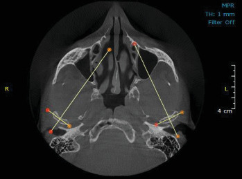
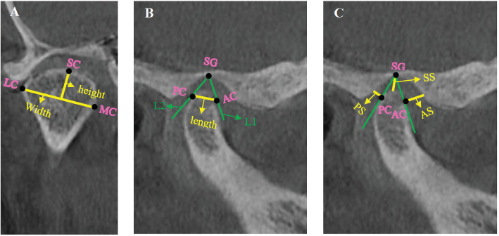
Purpose: The aim of this comparative observational study is to evaluate and compare the size and position of the condyle among male and female patients with different skeletal patterns in the anterior-posterior dimension using cone beam computed tomography (CBCT) images. Materials and Methods: CBCT images of 120 patients, all prepared for other treatment purposes under the same conditions, were included in the study. The patients were classified into three groups-class I, class II, and class III-based on ANB angles and Wits analysis. The size of the condyle was measured in terms of width, height, and length. The position of the condyle was assessed by measuring the superior joint space (SS), anterior joint space (AS), and posterior joint space (PS) on the right and left sides separately. The measurements and results were analyzed using analysis of covariance (ANCOVA) and Bonferroni analysis. A statistical significance level of p < 0.05 was considered. Results: The study found no statistically significant differences in the size of the SS and AS (p = 0.481 and p = 0.392, respectively) across different skeletal patterns. However, the size of the PS was significantly greater in class I subjects compared to class III subjects (p = 0.015). There were no statistically significant differences in condyle height and width among the different skeletal patterns (p = 0.367 and p = 0.720, respectively). In contrast, condyle length was statistically significant in class II individuals (p = 0.002) and was the lowest among the other skeletal pattern groups. Conclusions: Based on the results obtained, class I individuals have lower PS values compared to class III individuals. Additionally, class II individuals have shorter condyle lengths compared to those in class III and class I.


 分享
分享
 求助内容:
求助内容: 应助结果提醒方式:
 扫码关注我们
