Ultrasound localization microscopy in the diagnosis of breast tumors and prediction of relevant histologic biomarkers associated with prognosis in humans: the protocol for a prospective, multicenter study.
Jia Li, Lei Chen, Ronghui Wang, Jiang Zhu, Ao Li, Jianchun Li, Zhaojun Li, Wen Luo, Wenkun Bai, Tao Ying, Cong Wei, Di Sun, Yuanyi Zheng
{"title":"Ultrasound localization microscopy in the diagnosis of breast tumors and prediction of relevant histologic biomarkers associated with prognosis in humans: the protocol for a prospective, multicenter study.","authors":"Jia Li, Lei Chen, Ronghui Wang, Jiang Zhu, Ao Li, Jianchun Li, Zhaojun Li, Wen Luo, Wenkun Bai, Tao Ying, Cong Wei, Di Sun, Yuanyi Zheng","doi":"10.1186/s12880-024-01535-7","DOIUrl":null,"url":null,"abstract":"<p><strong>Background: </strong>Benign and malignant breast tumors differ in their microvasculature morphology and distribution. Histologic biomarkers of malignant breast tumors are also correlated with the microvasculature. There is a lack of imaging technology for evaluating the microvasculature. Ultrasound localization microscopy (ULM) can provide detailed microvascular architecture at super-resolution. The objective of this trial is to explore the role of ULM in distinguishing benign from malignant breast tumors and to explore the correlations between ULM qualitative and quantitative parameters and histologic biomarkers in malignant breast tumors.</p><p><strong>Methods/design: </strong>This prospective and multicenter study will include 83 patients with breast tumors that will undergo ULM. 55 patients will be assigned to the malignant group, and 28 patients will be assigned to the benign group. The primary outcome is the differences in the qualitative parameters (microvasculature morphology, distribution, and flow direction) between benign and malignant breast tumors on ULM. Secondary outcomes include (1) differences in the quantitative parameters (microvasculature density, tortuosity, diameter, and flow velocity) between benign and malignant breast tumors based on ULM; (2) diagnostic performance of the qualitative parameters in distinguishing benign and malignant breast tumors; (3) diagnostic performance of the quantitative parameters in distinguishing benign and malignant breast tumors; (4) relationships between the qualitative parameters and histologic biomarkers in malignant breast tumors; (5) relationships between the quantitative parameters and histologic biomarkers in malignant breast tumors; and (6) the evaluation of inter-reader and intra-reader reproducibility.</p><p><strong>Discussion: </strong>Detecting vascularity in breast tumors is of great significance to differentiate benign from malignant tumors and to predict histologic biomarkers. These histologic biomarkers, such as ER, PR, HER2 and Ki67, are closely related to prognosis evaluation. This trial will provide maximum information about the microvasculature of breast tumors and thereby will help with the formulation of subsequent differential diagnosis and the prediction of histologic biomarkers.</p><p><strong>Trial registration number/date: </strong>Chinese Clinical Trial Registry ChiCTR2100048361/6th/July/2021. This study is a part of that clinical trial.</p>","PeriodicalId":9020,"journal":{"name":"BMC Medical Imaging","volume":"25 1","pages":"13"},"PeriodicalIF":3.2000,"publicationDate":"2025-01-08","publicationTypes":"Journal Article","fieldsOfStudy":null,"isOpenAccess":false,"openAccessPdf":"https://www.ncbi.nlm.nih.gov/pmc/articles/PMC11715691/pdf/","citationCount":"0","resultStr":null,"platform":"Semanticscholar","paperid":null,"PeriodicalName":"BMC Medical Imaging","FirstCategoryId":"3","ListUrlMain":"https://doi.org/10.1186/s12880-024-01535-7","RegionNum":3,"RegionCategory":"医学","ArticlePicture":[],"TitleCN":null,"AbstractTextCN":null,"PMCID":null,"EPubDate":"","PubModel":"","JCR":"Q2","JCRName":"RADIOLOGY, NUCLEAR MEDICINE & MEDICAL IMAGING","Score":null,"Total":0}
引用次数: 0
Abstract
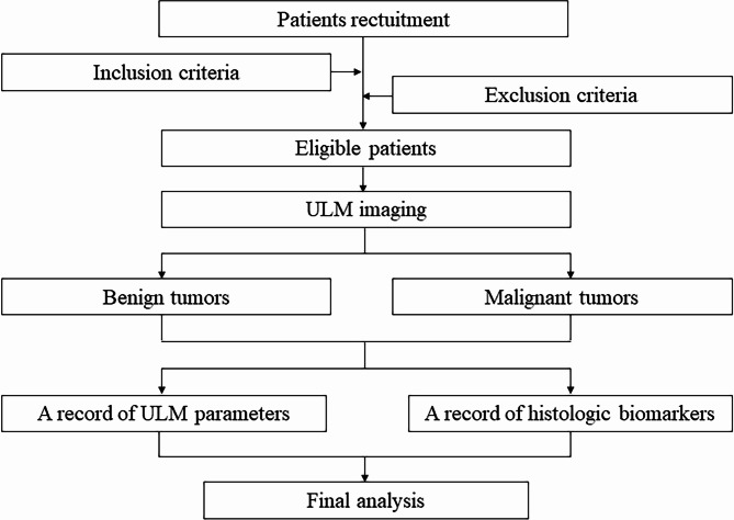
Background: Benign and malignant breast tumors differ in their microvasculature morphology and distribution. Histologic biomarkers of malignant breast tumors are also correlated with the microvasculature. There is a lack of imaging technology for evaluating the microvasculature. Ultrasound localization microscopy (ULM) can provide detailed microvascular architecture at super-resolution. The objective of this trial is to explore the role of ULM in distinguishing benign from malignant breast tumors and to explore the correlations between ULM qualitative and quantitative parameters and histologic biomarkers in malignant breast tumors.
Methods/design: This prospective and multicenter study will include 83 patients with breast tumors that will undergo ULM. 55 patients will be assigned to the malignant group, and 28 patients will be assigned to the benign group. The primary outcome is the differences in the qualitative parameters (microvasculature morphology, distribution, and flow direction) between benign and malignant breast tumors on ULM. Secondary outcomes include (1) differences in the quantitative parameters (microvasculature density, tortuosity, diameter, and flow velocity) between benign and malignant breast tumors based on ULM; (2) diagnostic performance of the qualitative parameters in distinguishing benign and malignant breast tumors; (3) diagnostic performance of the quantitative parameters in distinguishing benign and malignant breast tumors; (4) relationships between the qualitative parameters and histologic biomarkers in malignant breast tumors; (5) relationships between the quantitative parameters and histologic biomarkers in malignant breast tumors; and (6) the evaluation of inter-reader and intra-reader reproducibility.
Discussion: Detecting vascularity in breast tumors is of great significance to differentiate benign from malignant tumors and to predict histologic biomarkers. These histologic biomarkers, such as ER, PR, HER2 and Ki67, are closely related to prognosis evaluation. This trial will provide maximum information about the microvasculature of breast tumors and thereby will help with the formulation of subsequent differential diagnosis and the prediction of histologic biomarkers.
Trial registration number/date: Chinese Clinical Trial Registry ChiCTR2100048361/6th/July/2021. This study is a part of that clinical trial.
期刊介绍:
BMC Medical Imaging is an open access journal publishing original peer-reviewed research articles in the development, evaluation, and use of imaging techniques and image processing tools to diagnose and manage disease.


 分享
分享
 求助内容:
求助内容: 应助结果提醒方式:
 扫码关注我们
