Application value of CT three-dimensional reconstruction technology in the identification of benign and malignant lung nodules and the characteristics of nodule distribution.
Guanghai Ji, Fei Liu, Zhiqiang Chen, Jie Peng, Hao Deng, Sheng Xiao, Yun Li
{"title":"Application value of CT three-dimensional reconstruction technology in the identification of benign and malignant lung nodules and the characteristics of nodule distribution.","authors":"Guanghai Ji, Fei Liu, Zhiqiang Chen, Jie Peng, Hao Deng, Sheng Xiao, Yun Li","doi":"10.1186/s12880-024-01505-z","DOIUrl":null,"url":null,"abstract":"<p><strong>Objective: </strong>The study aimed to evaluate the application value of computed tomography (CT) three-dimensional (3D) reconstruction technology in identifying benign and malignant lung nodules and characterizing the distribution of the nodules.</p><p><strong>Methods: </strong>CT 3D reconstruction was performed for lung nodules. Pathological results were used as the gold standard to compare the detection rates of various lung nodule signs between conventional chest CT scanning and CT 3D reconstruction techniques. Additionally, the differences in mean diffusion coefficient values and partial anisotropy index values between male and female patients were analyzed.</p><p><strong>Results: </strong>Pathologic confirmation identified 30 patients with benign lesions and 45 patients with malignant lesions. CT 3D reconstruction demonstrated higher diagnostic accuracy for lung nodule imaging signs compared to conventional CT scanning (P < 0.05). The mean diffusion coefficient values and partial anisotropy index values were lower in female patients compared to male patients in the lung nodule lesion area, lung perinodular edema area, and normal lung tissue (P < 0.05). Conventional CT scanning showed a benign accuracy rate of 63.33% and a malignant accuracy rate of 60.00%, whereas CT 3D imaging achieved a benign and malignant accuracy rate of 86.67% for both. The accuracy rates for CT 3D imaging were significantly higher than those for conventional CT scanning (P < 0.05).</p><p><strong>Conclusion: </strong>CT 3D imaging technology demonstrates high diagnostic accuracy in differentiating benign from malignant lung nodules.</p>","PeriodicalId":9020,"journal":{"name":"BMC Medical Imaging","volume":"25 1","pages":"7"},"PeriodicalIF":3.2000,"publicationDate":"2025-01-06","publicationTypes":"Journal Article","fieldsOfStudy":null,"isOpenAccess":false,"openAccessPdf":"https://www.ncbi.nlm.nih.gov/pmc/articles/PMC11702159/pdf/","citationCount":"0","resultStr":null,"platform":"Semanticscholar","paperid":null,"PeriodicalName":"BMC Medical Imaging","FirstCategoryId":"3","ListUrlMain":"https://doi.org/10.1186/s12880-024-01505-z","RegionNum":3,"RegionCategory":"医学","ArticlePicture":[],"TitleCN":null,"AbstractTextCN":null,"PMCID":null,"EPubDate":"","PubModel":"","JCR":"Q2","JCRName":"RADIOLOGY, NUCLEAR MEDICINE & MEDICAL IMAGING","Score":null,"Total":0}
引用次数: 0
Abstract
Objective: The study aimed to evaluate the application value of computed tomography (CT) three-dimensional (3D) reconstruction technology in identifying benign and malignant lung nodules and characterizing the distribution of the nodules.
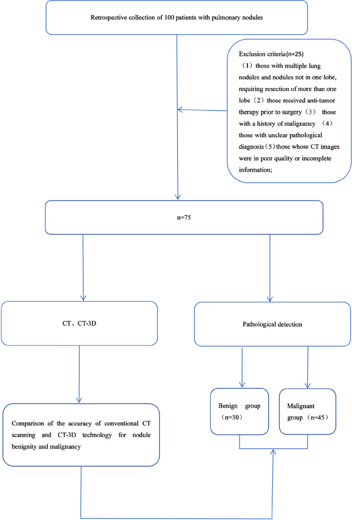
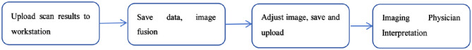
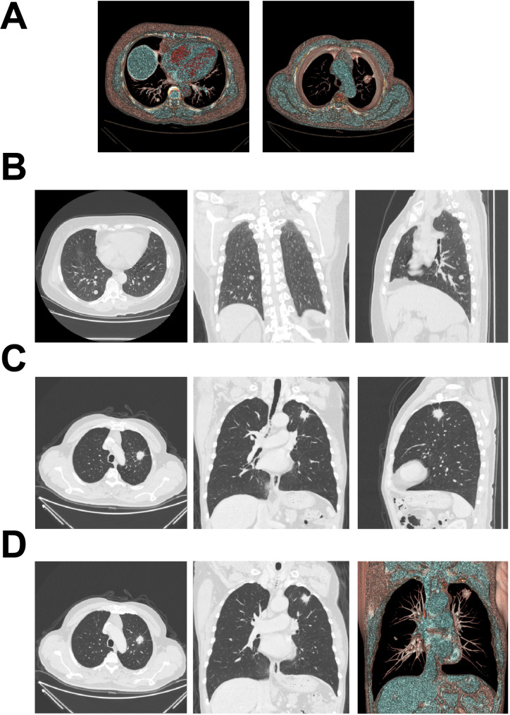
Methods: CT 3D reconstruction was performed for lung nodules. Pathological results were used as the gold standard to compare the detection rates of various lung nodule signs between conventional chest CT scanning and CT 3D reconstruction techniques. Additionally, the differences in mean diffusion coefficient values and partial anisotropy index values between male and female patients were analyzed.
Results: Pathologic confirmation identified 30 patients with benign lesions and 45 patients with malignant lesions. CT 3D reconstruction demonstrated higher diagnostic accuracy for lung nodule imaging signs compared to conventional CT scanning (P < 0.05). The mean diffusion coefficient values and partial anisotropy index values were lower in female patients compared to male patients in the lung nodule lesion area, lung perinodular edema area, and normal lung tissue (P < 0.05). Conventional CT scanning showed a benign accuracy rate of 63.33% and a malignant accuracy rate of 60.00%, whereas CT 3D imaging achieved a benign and malignant accuracy rate of 86.67% for both. The accuracy rates for CT 3D imaging were significantly higher than those for conventional CT scanning (P < 0.05).
Conclusion: CT 3D imaging technology demonstrates high diagnostic accuracy in differentiating benign from malignant lung nodules.
期刊介绍:
BMC Medical Imaging is an open access journal publishing original peer-reviewed research articles in the development, evaluation, and use of imaging techniques and image processing tools to diagnose and manage disease.


 分享
分享
 求助内容:
求助内容: 应助结果提醒方式:
 扫码关注我们
