Giuseppe Broggi, Jessica Farina, Valeria Barresi, Francesco Certo, Giuseppe Maria Vincenzo Barbagallo, Gaetano Magro, Rosario Caltabiano
{"title":"Immunohistochemical Expression of PAX8 in Central Nervous System Hemangioblastomas: A Potential Diagnostic Pitfall for Neuropathologists.","authors":"Giuseppe Broggi, Jessica Farina, Valeria Barresi, Francesco Certo, Giuseppe Maria Vincenzo Barbagallo, Gaetano Magro, Rosario Caltabiano","doi":"10.1097/PAI.0000000000001246","DOIUrl":null,"url":null,"abstract":"<p><p>The histologic differential diagnosis between intracranial hemangioblastoma (HB) and metastatic clear cell renal cell carcinoma may be challenging, especially considering that both tumors exhibit clear cell morphology and can be associated with vHL mutation and/or Von Hippel-Lindau syndrome. As the execution of immunohistochemical analyses is often mandatory, the expression of PAX8 has been traditionally considered a reliable marker of metastatic clear cell renal cell carcinoma, being consistently negative in intracranial HB. However, as in recent years, some cases of PAX8-positive HBs have been reported in the literature; we studied the expression of this antibody on a series of 23 intracranial HB, showing that about 40% of these tumors may express PAX8 and that this immunoreactivity is often focal and weak. We would like to emphasize that the possibility of a PAX8-positive intracranial HB does exist and must be taken into account by neuropathologists to avoid misdiagnoses; in this regard, a broader immunohistochemical panel also including CD10, Inhibin-α, PAX2, S100, and anti-Renal cell carcinoma (RCC) antibody is highly recommended.</p>","PeriodicalId":48952,"journal":{"name":"Applied Immunohistochemistry & Molecular Morphology","volume":" ","pages":"160-163"},"PeriodicalIF":1.2000,"publicationDate":"2025-05-01","publicationTypes":"Journal Article","fieldsOfStudy":null,"isOpenAccess":false,"openAccessPdf":"https://www.ncbi.nlm.nih.gov/pmc/articles/PMC12043255/pdf/","citationCount":"0","resultStr":null,"platform":"Semanticscholar","paperid":null,"PeriodicalName":"Applied Immunohistochemistry & Molecular Morphology","FirstCategoryId":"3","ListUrlMain":"https://doi.org/10.1097/PAI.0000000000001246","RegionNum":4,"RegionCategory":"医学","ArticlePicture":[],"TitleCN":null,"AbstractTextCN":null,"PMCID":null,"EPubDate":"2025/1/27 0:00:00","PubModel":"Epub","JCR":"Q3","JCRName":"ANATOMY & MORPHOLOGY","Score":null,"Total":0}
引用次数: 0
Abstract
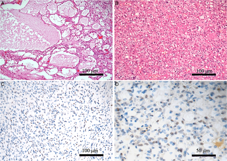
The histologic differential diagnosis between intracranial hemangioblastoma (HB) and metastatic clear cell renal cell carcinoma may be challenging, especially considering that both tumors exhibit clear cell morphology and can be associated with vHL mutation and/or Von Hippel-Lindau syndrome. As the execution of immunohistochemical analyses is often mandatory, the expression of PAX8 has been traditionally considered a reliable marker of metastatic clear cell renal cell carcinoma, being consistently negative in intracranial HB. However, as in recent years, some cases of PAX8-positive HBs have been reported in the literature; we studied the expression of this antibody on a series of 23 intracranial HB, showing that about 40% of these tumors may express PAX8 and that this immunoreactivity is often focal and weak. We would like to emphasize that the possibility of a PAX8-positive intracranial HB does exist and must be taken into account by neuropathologists to avoid misdiagnoses; in this regard, a broader immunohistochemical panel also including CD10, Inhibin-α, PAX2, S100, and anti-Renal cell carcinoma (RCC) antibody is highly recommended.
期刊介绍:
Applied Immunohistochemistry & Molecular Morphology covers newly developed identification and detection technologies, and their applications in research and diagnosis for the applied immunohistochemist & molecular Morphologist.
Official Journal of the International Society for Immunohistochemisty and Molecular Morphology.


 分享
分享
 求助内容:
求助内容: 应助结果提醒方式:
 扫码关注我们
