Sebastian Kenny, Shalini Iyer, Clinton A. Gabel, Natalia Tegenfeldt, Andrew G. DeMarco, Mark C. Hall, Leifu Chang, V. Jo Davisson, Scott Vande Pol, Chittaranjan Das
{"title":"Structure of E6AP in complex with HPV16-E6 and p53 reveals a novel ordered domain important for E3 ligase activation","authors":"Sebastian Kenny, Shalini Iyer, Clinton A. Gabel, Natalia Tegenfeldt, Andrew G. DeMarco, Mark C. Hall, Leifu Chang, V. Jo Davisson, Scott Vande Pol, Chittaranjan Das","doi":"10.1016/j.str.2024.12.013","DOIUrl":null,"url":null,"abstract":"High-risk human papillomavirus E6 oncoprotein is a model system for the recognition and degradation of cellular p53 tumor suppressor protein. There remains a gap in the understanding of the ubiquitin transfer reaction, including placement of the E6AP catalytic HECT domain of the ligase concerning the p53 substrate and how E6 itself is protected from ubiquitination. We determined the cryoelectron microscopy (cryo-EM) structure of the E6AP/E6/p53 complex, related the structure to <em>in vivo</em> modeling of the tri-molecular complex, and identified structural interactions associated with activation of the ubiquitin ligase function. The structure reveals that the N-terminal ordered domain (NOD) in E6AP has a terminal alpha helix that mediates the interaction of the NOD with the HECT domain of E6AP and protects the HPV-E6 protein from ubiquitination. In addition, this NOD helix is required for E6AP ligase function by contributing to the affinity of the E6-E6AP association, modulating E6 substrate recognition, while displacing UbcH7.","PeriodicalId":22168,"journal":{"name":"Structure","volume":"41 1","pages":""},"PeriodicalIF":4.3000,"publicationDate":"2025-01-15","publicationTypes":"Journal Article","fieldsOfStudy":null,"isOpenAccess":false,"openAccessPdf":"","citationCount":"0","resultStr":null,"platform":"Semanticscholar","paperid":null,"PeriodicalName":"Structure","FirstCategoryId":"99","ListUrlMain":"https://doi.org/10.1016/j.str.2024.12.013","RegionNum":2,"RegionCategory":"生物学","ArticlePicture":[],"TitleCN":null,"AbstractTextCN":null,"PMCID":null,"EPubDate":"","PubModel":"","JCR":"Q2","JCRName":"BIOCHEMISTRY & MOLECULAR BIOLOGY","Score":null,"Total":0}
引用次数: 0
Abstract
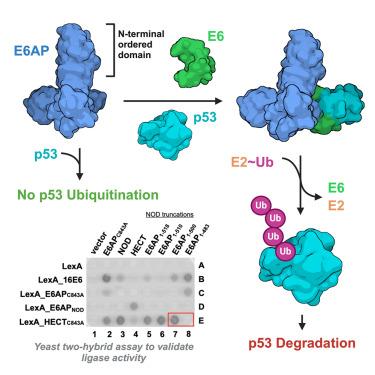
High-risk human papillomavirus E6 oncoprotein is a model system for the recognition and degradation of cellular p53 tumor suppressor protein. There remains a gap in the understanding of the ubiquitin transfer reaction, including placement of the E6AP catalytic HECT domain of the ligase concerning the p53 substrate and how E6 itself is protected from ubiquitination. We determined the cryoelectron microscopy (cryo-EM) structure of the E6AP/E6/p53 complex, related the structure to in vivo modeling of the tri-molecular complex, and identified structural interactions associated with activation of the ubiquitin ligase function. The structure reveals that the N-terminal ordered domain (NOD) in E6AP has a terminal alpha helix that mediates the interaction of the NOD with the HECT domain of E6AP and protects the HPV-E6 protein from ubiquitination. In addition, this NOD helix is required for E6AP ligase function by contributing to the affinity of the E6-E6AP association, modulating E6 substrate recognition, while displacing UbcH7.
期刊介绍:
Structure aims to publish papers of exceptional interest in the field of structural biology. The journal strives to be essential reading for structural biologists, as well as biologists and biochemists that are interested in macromolecular structure and function. Structure strongly encourages the submission of manuscripts that present structural and molecular insights into biological function and mechanism. Other reports that address fundamental questions in structural biology, such as structure-based examinations of protein evolution, folding, and/or design, will also be considered. We will consider the application of any method, experimental or computational, at high or low resolution, to conduct structural investigations, as long as the method is appropriate for the biological, functional, and mechanistic question(s) being addressed. Likewise, reports describing single-molecule analysis of biological mechanisms are welcome.
In general, the editors encourage submission of experimental structural studies that are enriched by an analysis of structure-activity relationships and will not consider studies that solely report structural information unless the structure or analysis is of exceptional and broad interest. Studies reporting only homology models, de novo models, or molecular dynamics simulations are also discouraged unless the models are informed by or validated by novel experimental data; rationalization of a large body of existing experimental evidence and making testable predictions based on a model or simulation is often not considered sufficient.


 分享
分享
 求助内容:
求助内容: 应助结果提醒方式:
 扫码关注我们
