Matthias Witt, Uli Weber, Sebastian Adeberg, Kilian-Simon Baumann, Klemens Zink
{"title":"Proton and Carbon Ion Beam Spot Size Measurement Using 5 Different Detector Types.","authors":"Matthias Witt, Uli Weber, Sebastian Adeberg, Kilian-Simon Baumann, Klemens Zink","doi":"10.1016/j.ijpt.2024.100638","DOIUrl":null,"url":null,"abstract":"<p><strong>Purpose: </strong>The spot size of scanned particle beams is of crucial importance for the correct dose delivery and, therefore, plays a significant role in the quality assurance (QA) of pencil beam scanning ion beam therapy.</p><p><strong>Materials and methods: </strong>This study compares 5 detector types-radiochromic film, ionization chamber (IC) array, flat panel detector, multiwire chamber, and IC-for measuring the spot size of proton and carbon ion beams.</p><p><strong>Results: </strong>Variations of up to 30% were found between detectors, underscoring the impact of detector choice on QA outcomes. The multiwire chamber consistently measured the smallest spot sizes, attributed to its intrinsic calculation model, while the IC array yielded larger spot sizes due to volume-averaging effects. These discrepancies highlight the necessity of selecting detectors based on QA needs, such as measurement speed, spatial resolution, and data acquisition methods. Digital detectors offer advantages over film-based ones by automating data processing, reducing manual errors, and providing immediate results.</p><p><strong>Conclusion: </strong>The study concludes that, although a single Gaussian fit is generally sufficient for QA, more sophisticated models might be beneficial for special applications. These findings aim to guide detector selection for ion beam facilities, enhancing QA procedures.</p>","PeriodicalId":36923,"journal":{"name":"International Journal of Particle Therapy","volume":"15 ","pages":"100638"},"PeriodicalIF":2.0000,"publicationDate":"2024-12-13","publicationTypes":"Journal Article","fieldsOfStudy":null,"isOpenAccess":false,"openAccessPdf":"https://www.ncbi.nlm.nih.gov/pmc/articles/PMC11732072/pdf/","citationCount":"0","resultStr":null,"platform":"Semanticscholar","paperid":null,"PeriodicalName":"International Journal of Particle Therapy","FirstCategoryId":"1085","ListUrlMain":"https://doi.org/10.1016/j.ijpt.2024.100638","RegionNum":0,"RegionCategory":null,"ArticlePicture":[],"TitleCN":null,"AbstractTextCN":null,"PMCID":null,"EPubDate":"2025/3/1 0:00:00","PubModel":"eCollection","JCR":"Q3","JCRName":"ONCOLOGY","Score":null,"Total":0}
引用次数: 0
Abstract
Purpose: The spot size of scanned particle beams is of crucial importance for the correct dose delivery and, therefore, plays a significant role in the quality assurance (QA) of pencil beam scanning ion beam therapy.
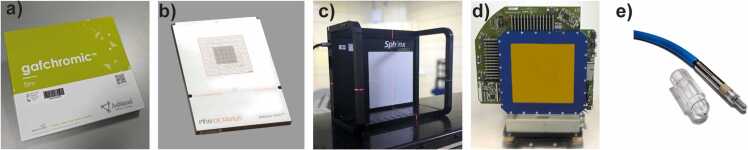
Materials and methods: This study compares 5 detector types-radiochromic film, ionization chamber (IC) array, flat panel detector, multiwire chamber, and IC-for measuring the spot size of proton and carbon ion beams.
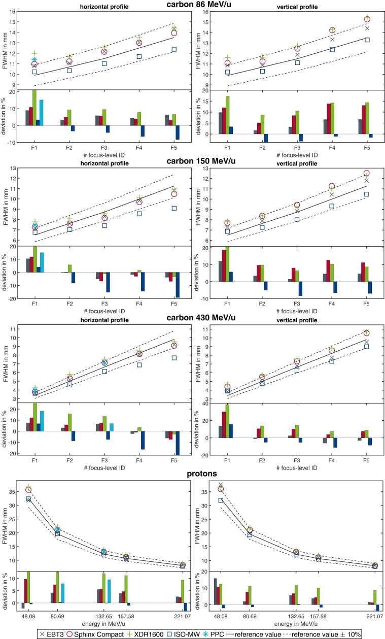
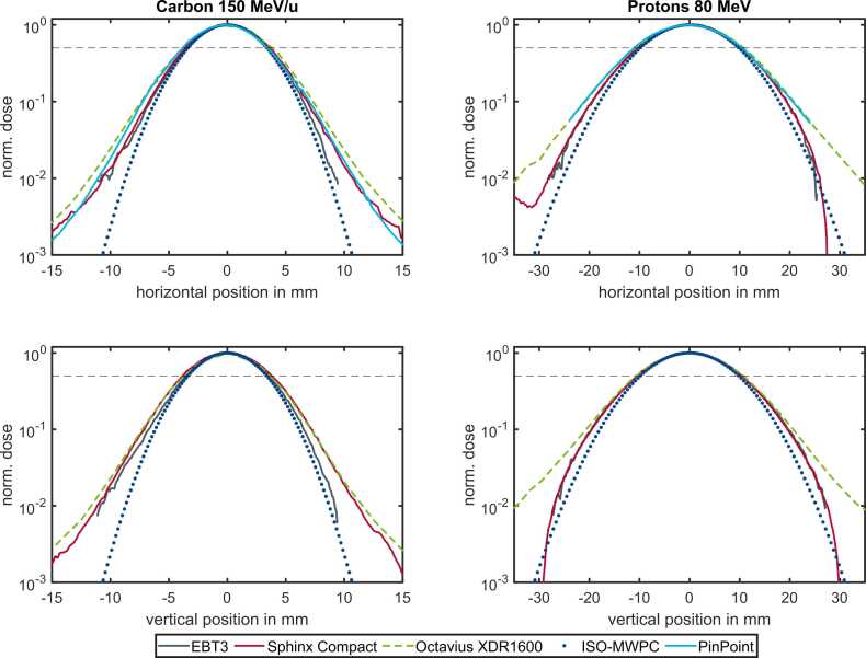
Results: Variations of up to 30% were found between detectors, underscoring the impact of detector choice on QA outcomes. The multiwire chamber consistently measured the smallest spot sizes, attributed to its intrinsic calculation model, while the IC array yielded larger spot sizes due to volume-averaging effects. These discrepancies highlight the necessity of selecting detectors based on QA needs, such as measurement speed, spatial resolution, and data acquisition methods. Digital detectors offer advantages over film-based ones by automating data processing, reducing manual errors, and providing immediate results.
Conclusion: The study concludes that, although a single Gaussian fit is generally sufficient for QA, more sophisticated models might be beneficial for special applications. These findings aim to guide detector selection for ion beam facilities, enhancing QA procedures.


 分享
分享
 求助内容:
求助内容: 应助结果提醒方式:
 扫码关注我们
