{"title":"Clinical study of colorViz fusion image vascular grading based on multi-phase CTA reconstruction in acute ischemic stroke.","authors":"Qi Wang, Qiang Wang, Yunfa Xu, Xue Li, Dingbin Zhou, Xiaotong Sun, Bo Feng","doi":"10.1186/s12880-024-01490-3","DOIUrl":null,"url":null,"abstract":"<p><strong>Objective: </strong>This study aimed to evaluate the diagnostic value of ColorViz fused images from multi-phase computed tomography angiography (mCTA) using GE Healthcare's FastStroke software for newly diagnosed cerebral infarctions in patients with acute ischemic stroke (AIS).</p><p><strong>Methods: </strong>A total of 106 AIS patients with unilateral anterior circulation occlusion were prospectively enrolled. All patients underwent mCTA scans during the arterial peak phase, venous peak phase, and venous late phase. The vascular information from these mCTA phases was combined into a time-varying color-coded image using GE Healthcare's FastStroke software. All participants also underwent magnetic resonance diffusion-weighted imaging (MR-DWI) within three days. The diagnostic capability of the mCTA ColorViz fusion images for identifying newly diagnosed intracranial infarction was assessed using MR-DWI as the gold standard, focusing on the degree of delayed vascular perfusion and the number of visible blood vessels.</p><p><strong>Results: </strong>The mCTA ColorViz fusion images revealed ischemic changes in brain tissue, demonstrating a sensitivity of 88.7% for superficial infarctions and 48.5% for deep infarctions. Additionally, the subjective vascular grading score of the mCTA ColorViz fusion images showed a strong negative correlation with the infarct area identified by MR-DWI (r = - 0.6, P < 0.001).</p><p><strong>Conclusion: </strong>The mCTA ColorViz fusion images produced by FastStroke software provide valuable diagnostic insights for newly diagnosed cerebral infarction in AIS patients. The sensitivity of these images is notably higher for superficial infarctions compared to deep ones. This technique allows for relatively accurate detection of the ischemic extent and the likelihood of infarction in the superficial regions where lesions are located.</p>","PeriodicalId":9020,"journal":{"name":"BMC Medical Imaging","volume":"25 1","pages":"25"},"PeriodicalIF":3.2000,"publicationDate":"2025-01-21","publicationTypes":"Journal Article","fieldsOfStudy":null,"isOpenAccess":false,"openAccessPdf":"https://www.ncbi.nlm.nih.gov/pmc/articles/PMC11748880/pdf/","citationCount":"0","resultStr":null,"platform":"Semanticscholar","paperid":null,"PeriodicalName":"BMC Medical Imaging","FirstCategoryId":"3","ListUrlMain":"https://doi.org/10.1186/s12880-024-01490-3","RegionNum":3,"RegionCategory":"医学","ArticlePicture":[],"TitleCN":null,"AbstractTextCN":null,"PMCID":null,"EPubDate":"","PubModel":"","JCR":"Q2","JCRName":"RADIOLOGY, NUCLEAR MEDICINE & MEDICAL IMAGING","Score":null,"Total":0}
引用次数: 0
Abstract
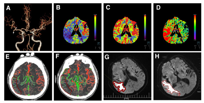
Objective: This study aimed to evaluate the diagnostic value of ColorViz fused images from multi-phase computed tomography angiography (mCTA) using GE Healthcare's FastStroke software for newly diagnosed cerebral infarctions in patients with acute ischemic stroke (AIS).
Methods: A total of 106 AIS patients with unilateral anterior circulation occlusion were prospectively enrolled. All patients underwent mCTA scans during the arterial peak phase, venous peak phase, and venous late phase. The vascular information from these mCTA phases was combined into a time-varying color-coded image using GE Healthcare's FastStroke software. All participants also underwent magnetic resonance diffusion-weighted imaging (MR-DWI) within three days. The diagnostic capability of the mCTA ColorViz fusion images for identifying newly diagnosed intracranial infarction was assessed using MR-DWI as the gold standard, focusing on the degree of delayed vascular perfusion and the number of visible blood vessels.
Results: The mCTA ColorViz fusion images revealed ischemic changes in brain tissue, demonstrating a sensitivity of 88.7% for superficial infarctions and 48.5% for deep infarctions. Additionally, the subjective vascular grading score of the mCTA ColorViz fusion images showed a strong negative correlation with the infarct area identified by MR-DWI (r = - 0.6, P < 0.001).
Conclusion: The mCTA ColorViz fusion images produced by FastStroke software provide valuable diagnostic insights for newly diagnosed cerebral infarction in AIS patients. The sensitivity of these images is notably higher for superficial infarctions compared to deep ones. This technique allows for relatively accurate detection of the ischemic extent and the likelihood of infarction in the superficial regions where lesions are located.
期刊介绍:
BMC Medical Imaging is an open access journal publishing original peer-reviewed research articles in the development, evaluation, and use of imaging techniques and image processing tools to diagnose and manage disease.


 分享
分享
 求助内容:
求助内容: 应助结果提醒方式:
 扫码关注我们
