{"title":"Parietal Meningocele Under the Scalp of a Fetus Diagnosed Based on Volume Contrast Imaging of Prenatal Three-Dimensional Ultrasound Data.","authors":"Akihiro Hasegawa, Masami Kono, Tokumasa Suemitsu, Yuki Ito, Tatsuya Hirotsu, Yuichiro Nonaka, Osamu Samura, Aikou Okamoto","doi":"10.1155/crnm/7401673","DOIUrl":null,"url":null,"abstract":"<p><p>Determining the differential diagnosis of small scalp cysts identified on a fetus is difficult. In particular, many physicians have difficulty differentiating small meningoceles from small scalp cysts during the prenatal period. Volume contrast imaging increases contrast between tissues, thereby allowing an enhanced view of target structures. A 15 × 5 mm scalp cyst was identified on a fetus during a prenatal ultrasonography examination performed at 20 weeks of gestation. The cyst was not connected to the blood flow, and did not include the tissue of the brain parenchyma. Ventriculomegaly and other structural abnormalities were not observed. Based on these findings, we suspected a sinus pericranii or fetal epidermal cyst. The size of the fetal scalp cyst was stable, and the growth of the fetus remained normal until birth. The diagnosis of a small meningocele was confirmed postnatally, based on the results of a magnetic resonance imaging examination. Postnatal evaluation of offline volume contrast imaging of prenatal three-dimensional ultrasound data at 22 weeks of gestation revealed a skull bone defect beneath the cyst. Volume contrast imaging can facilitate the prenatal diagnosis of small meningoceles by detecting bone defects on the fetal head.</p>","PeriodicalId":9615,"journal":{"name":"Case Reports in Neurological Medicine","volume":"2025 ","pages":"7401673"},"PeriodicalIF":0.9000,"publicationDate":"2025-01-08","publicationTypes":"Journal Article","fieldsOfStudy":null,"isOpenAccess":false,"openAccessPdf":"https://www.ncbi.nlm.nih.gov/pmc/articles/PMC11735054/pdf/","citationCount":"0","resultStr":null,"platform":"Semanticscholar","paperid":null,"PeriodicalName":"Case Reports in Neurological Medicine","FirstCategoryId":"1085","ListUrlMain":"https://doi.org/10.1155/crnm/7401673","RegionNum":0,"RegionCategory":null,"ArticlePicture":[],"TitleCN":null,"AbstractTextCN":null,"PMCID":null,"EPubDate":"2025/1/1 0:00:00","PubModel":"eCollection","JCR":"Q4","JCRName":"CLINICAL NEUROLOGY","Score":null,"Total":0}
引用次数: 0
Abstract
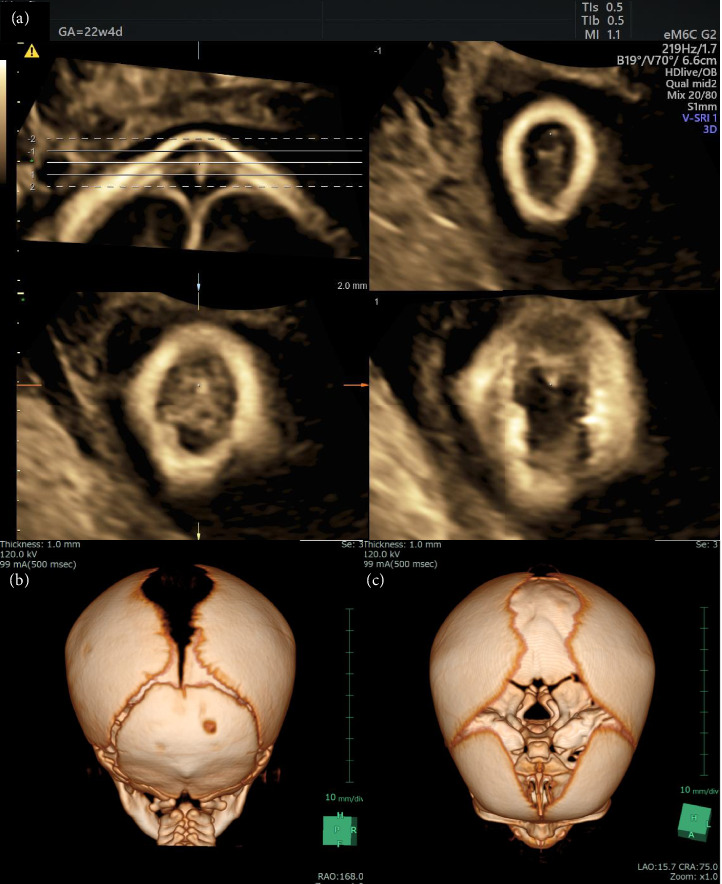
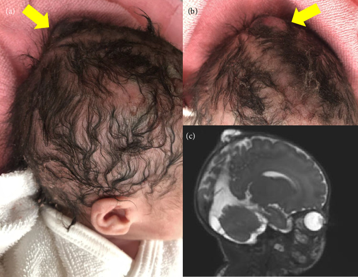
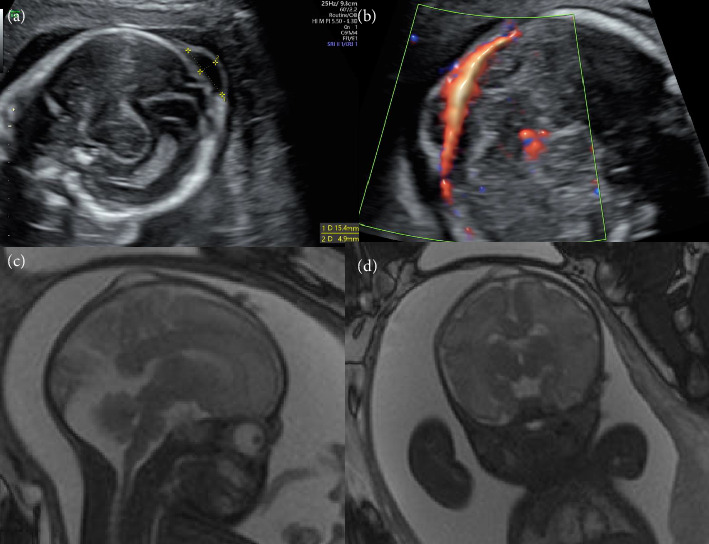
Determining the differential diagnosis of small scalp cysts identified on a fetus is difficult. In particular, many physicians have difficulty differentiating small meningoceles from small scalp cysts during the prenatal period. Volume contrast imaging increases contrast between tissues, thereby allowing an enhanced view of target structures. A 15 × 5 mm scalp cyst was identified on a fetus during a prenatal ultrasonography examination performed at 20 weeks of gestation. The cyst was not connected to the blood flow, and did not include the tissue of the brain parenchyma. Ventriculomegaly and other structural abnormalities were not observed. Based on these findings, we suspected a sinus pericranii or fetal epidermal cyst. The size of the fetal scalp cyst was stable, and the growth of the fetus remained normal until birth. The diagnosis of a small meningocele was confirmed postnatally, based on the results of a magnetic resonance imaging examination. Postnatal evaluation of offline volume contrast imaging of prenatal three-dimensional ultrasound data at 22 weeks of gestation revealed a skull bone defect beneath the cyst. Volume contrast imaging can facilitate the prenatal diagnosis of small meningoceles by detecting bone defects on the fetal head.


 分享
分享
 求助内容:
求助内容: 应助结果提醒方式:
 扫码关注我们
