{"title":"Development of Digestive Tract in Larval and Juvenile Red Spotted Grouper, <i>Epinephelus akaara</i>.","authors":"Moon-Soo Boo, Chi-Hoon Lee, Young-Don Lee","doi":"10.12717/DR.2024.28.4.187","DOIUrl":null,"url":null,"abstract":"<p><p>This study investigated the progressive morphological alterations and digestive tract development in larval and juvenile red spotted grouper, <i>Epinephelus akaara</i> across growth stages. External shape observations were made using an optical microscope, and the development of the digestive tract was investigated using histological methods. At 1 day after hatching (DAH), the digestive tract appeared as a straight tube extending between the ventral side and yolk-sac. At 3 DAH, the yolk was nearly absorbed, liver and pancreatic tissue began to develop. At 5 DAH, the opening of the mouth and anus allowed for the ingestion of external food, and the expansion of the intestinal lumen was observed. Gastric lumen differentiation became evident between the anterior intestine and esophagus. At 10 DAH, mucosal folds had formed in the rectum, and goblet cells were observed in the esophagus. At 20 DAH, mucosal folds in the stomach developed, and an increase in goblet cells was observed throughout the digestive organs. At 30 DAH, pyloric caeca and further gastric development were observed. By 40-50 DAH, sphincters between the esophagus, stomach, and intestine were clearly defined, resembling the adult digestive system. These findings suggest transitioning larvae to commercial pellets around 30 DAH, coinciding with the development of gastric glands and pyloric caeca. This research provides critical insights for optimizing feeding schedules to improve growth and survival rates during red spotted grouper seed production.</p>","PeriodicalId":72791,"journal":{"name":"Development & reproduction","volume":"28 4","pages":"187-194"},"PeriodicalIF":0.0000,"publicationDate":"2024-12-01","publicationTypes":"Journal Article","fieldsOfStudy":null,"isOpenAccess":false,"openAccessPdf":"https://www.ncbi.nlm.nih.gov/pmc/articles/PMC11750166/pdf/","citationCount":"0","resultStr":null,"platform":"Semanticscholar","paperid":null,"PeriodicalName":"Development & reproduction","FirstCategoryId":"1085","ListUrlMain":"https://doi.org/10.12717/DR.2024.28.4.187","RegionNum":0,"RegionCategory":null,"ArticlePicture":[],"TitleCN":null,"AbstractTextCN":null,"PMCID":null,"EPubDate":"2024/12/31 0:00:00","PubModel":"Epub","JCR":"","JCRName":"","Score":null,"Total":0}
引用次数: 0
Abstract
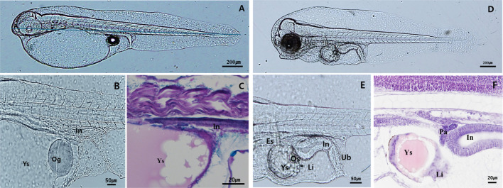
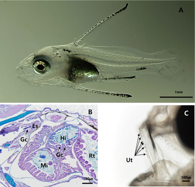
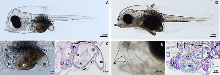
This study investigated the progressive morphological alterations and digestive tract development in larval and juvenile red spotted grouper, Epinephelus akaara across growth stages. External shape observations were made using an optical microscope, and the development of the digestive tract was investigated using histological methods. At 1 day after hatching (DAH), the digestive tract appeared as a straight tube extending between the ventral side and yolk-sac. At 3 DAH, the yolk was nearly absorbed, liver and pancreatic tissue began to develop. At 5 DAH, the opening of the mouth and anus allowed for the ingestion of external food, and the expansion of the intestinal lumen was observed. Gastric lumen differentiation became evident between the anterior intestine and esophagus. At 10 DAH, mucosal folds had formed in the rectum, and goblet cells were observed in the esophagus. At 20 DAH, mucosal folds in the stomach developed, and an increase in goblet cells was observed throughout the digestive organs. At 30 DAH, pyloric caeca and further gastric development were observed. By 40-50 DAH, sphincters between the esophagus, stomach, and intestine were clearly defined, resembling the adult digestive system. These findings suggest transitioning larvae to commercial pellets around 30 DAH, coinciding with the development of gastric glands and pyloric caeca. This research provides critical insights for optimizing feeding schedules to improve growth and survival rates during red spotted grouper seed production.


 分享
分享

 求助内容:
求助内容: 应助结果提醒方式:
 扫码关注我们
