{"title":"Prediction of isocitrate dehydrogenase mutation status in WHO grade II glioma by diffusion kurtosis imaging.","authors":"Wenjie Wu, Hui Zhang","doi":"10.5114/pjr/195521","DOIUrl":null,"url":null,"abstract":"<p><strong>Purpose: </strong>Isocitrate dehydrogenase (IDH) mutation status serves as a crucial prognostic indicator for glioma, typically assessed via immunohistochemical analysis post-surgery. Given the invasiveness of this approach, perhaps we can utilise convenient and noninvasive magnetic resonance imaging (MRI) methods to predict IDH mutation status. However, the current landscape lacks a standardised MRI technique for accurately predicting IDH mutations. In this study, we explore the potential of MRI diffusion kurtosis imaging (DKI) in forecasting the IDH mutation status of WHO grade II brain gliomas.</p><p><strong>Material and methods: </strong>Twenty-five patients with WHO grade II gliomas were retrospectively included. Patients underwent routine MRI and DKI scanning before surgery, measuring tumoural solid portion, peritumoral oedema, and normal-appearing white matter (NAWM) DKI parameters, including fractional anisotropy (FA), mean diffusivity (MD), mean kurtosis (MK), axial kurtosis (Ka), and axial radial kurtosis (Kr). The DKI parameter corrections were made (tumour or oedema parameters values divided by the NAWM value) to obtain the rFA (ratio of FA), rMD (ratio of MD), rMK (ratio of MK), rKA (ratio of KA), and rKr (ratio of Kr) values. Postoperative specimens were made of wax blocks and analysed by Sanger gene sequencing. DKI parameters between the 2 groups were compared by independent sample <i>t</i>-tests. The ROC curve was used to analyse the diagnostic value of each parameter.</p><p><strong>Results: </strong>Twenty-five patients were diagnosed with IDH-mutant (16 cases) and IDH-wild type (9 cases). The rFA and rMK values in the parenchymal region of IDH wild-type tumour were higher than those of IDH mutant, while the rMD values were lower than those of IDH mutant, and the difference between them was statistically significant (<i>p</i> < 0.05). The values of DKI parameters of peritumoral oedema in the 2 groups were not statistically significant.</p><p><strong>Conclusions: </strong>DKI can provide microstructural changes of diseased tissues and provide more imaging information for preoperative non-invasive judgment of IDH mutation status of WHO grade II gliomas. The values of rMK, rFA, and rMD are helpful in the assessment IDH mutation status, benefiting accurate diagnoses and treatment decisions.</p>","PeriodicalId":94174,"journal":{"name":"Polish journal of radiology","volume":"89 ","pages":"e566-e572"},"PeriodicalIF":0.0000,"publicationDate":"2024-12-20","publicationTypes":"Journal Article","fieldsOfStudy":null,"isOpenAccess":false,"openAccessPdf":"https://www.ncbi.nlm.nih.gov/pmc/articles/PMC11756366/pdf/","citationCount":"0","resultStr":null,"platform":"Semanticscholar","paperid":null,"PeriodicalName":"Polish journal of radiology","FirstCategoryId":"1085","ListUrlMain":"https://doi.org/10.5114/pjr/195521","RegionNum":0,"RegionCategory":null,"ArticlePicture":[],"TitleCN":null,"AbstractTextCN":null,"PMCID":null,"EPubDate":"2024/1/1 0:00:00","PubModel":"eCollection","JCR":"","JCRName":"","Score":null,"Total":0}
引用次数: 0
Abstract
Purpose: Isocitrate dehydrogenase (IDH) mutation status serves as a crucial prognostic indicator for glioma, typically assessed via immunohistochemical analysis post-surgery. Given the invasiveness of this approach, perhaps we can utilise convenient and noninvasive magnetic resonance imaging (MRI) methods to predict IDH mutation status. However, the current landscape lacks a standardised MRI technique for accurately predicting IDH mutations. In this study, we explore the potential of MRI diffusion kurtosis imaging (DKI) in forecasting the IDH mutation status of WHO grade II brain gliomas.
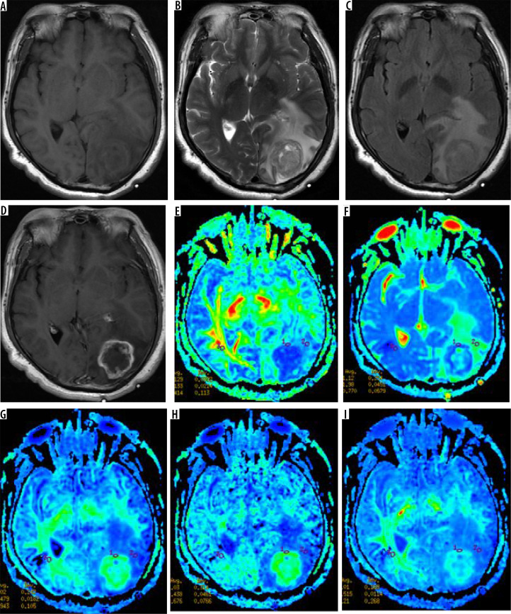
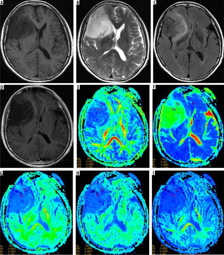
Material and methods: Twenty-five patients with WHO grade II gliomas were retrospectively included. Patients underwent routine MRI and DKI scanning before surgery, measuring tumoural solid portion, peritumoral oedema, and normal-appearing white matter (NAWM) DKI parameters, including fractional anisotropy (FA), mean diffusivity (MD), mean kurtosis (MK), axial kurtosis (Ka), and axial radial kurtosis (Kr). The DKI parameter corrections were made (tumour or oedema parameters values divided by the NAWM value) to obtain the rFA (ratio of FA), rMD (ratio of MD), rMK (ratio of MK), rKA (ratio of KA), and rKr (ratio of Kr) values. Postoperative specimens were made of wax blocks and analysed by Sanger gene sequencing. DKI parameters between the 2 groups were compared by independent sample t-tests. The ROC curve was used to analyse the diagnostic value of each parameter.
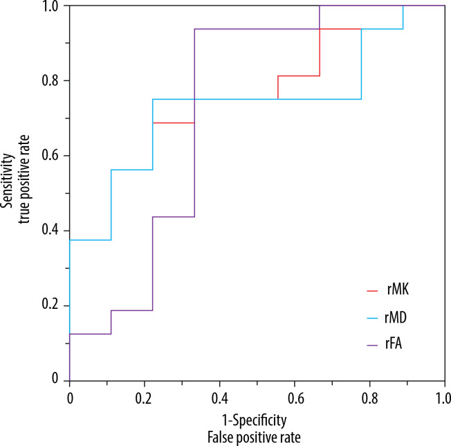
Results: Twenty-five patients were diagnosed with IDH-mutant (16 cases) and IDH-wild type (9 cases). The rFA and rMK values in the parenchymal region of IDH wild-type tumour were higher than those of IDH mutant, while the rMD values were lower than those of IDH mutant, and the difference between them was statistically significant (p < 0.05). The values of DKI parameters of peritumoral oedema in the 2 groups were not statistically significant.
Conclusions: DKI can provide microstructural changes of diseased tissues and provide more imaging information for preoperative non-invasive judgment of IDH mutation status of WHO grade II gliomas. The values of rMK, rFA, and rMD are helpful in the assessment IDH mutation status, benefiting accurate diagnoses and treatment decisions.


 分享
分享

 求助内容:
求助内容: 应助结果提醒方式:
 扫码关注我们
