{"title":"Deep learning system for the differential diagnosis of oral mucosal lesions through clinical photographic imaging","authors":"An-Yu Su , Ming-Long Wu , Yu-Hsueh Wu","doi":"10.1016/j.jds.2024.10.019","DOIUrl":null,"url":null,"abstract":"<div><h3>Background/purpose</h3><div>Oral mucosal lesions are associated with a variety of pathological conditions. Most deep-learning-based convolutional neural network (CNN) systems for computer-aided diagnosis of oral lesions have typically concentrated on determining limited aspects of differential diagnosis. This study aimed to develop a CNN-based diagnostic model capable of classifying clinical photographs of oral ulcerative and associated lesions into five different diagnoses, thereby assisting clinicians in making accurate differential diagnoses.</div></div><div><h3>Materials and methods</h3><div>A set of clinical images were selected, including 506 images of five different diagnoses. The images were pre-processed and randomly divided into two sets for training and testing the CNN model. The model architecture was composed of convolutional layers, batch normalization layers, max pooling layers, the dropout layer and fully-connected layers. Evaluation metrics included weighted-precision, weighted-recall, weighted-F1 score, average specificity, Cohen’s Kappa coefficient, normalized confusion matrix and AUC.</div></div><div><h3>Results</h3><div>The overall performance for the image classification showed a weighted-precision of 88.8%, a weighted-recall of 88.2%, a weighted-F1 score of 0.878, an average pecificity of 97.0%, a Kappa coefficient of 0.851, and an average AUC of 0.985.</div></div><div><h3>Conclusion</h3><div>The model achieved a decent classification performance (overall AUC=0.985), showing the capacity to discern between benign and malignant potential lesions, and laid the foundation of a novel tool that can help clinical differential diagnosis of oral mucosal lesions. The main challenges were the small and imbalanced dataset. Enlarging the minority classes, incorporating more oral mucosal lesion diagnoses, employing transfer learning and cross-validation might be included in future works to optimize the image classification model.</div></div>","PeriodicalId":15583,"journal":{"name":"Journal of Dental Sciences","volume":"20 1","pages":"Pages 54-60"},"PeriodicalIF":3.1000,"publicationDate":"2025-01-01","publicationTypes":"Journal Article","fieldsOfStudy":null,"isOpenAccess":false,"openAccessPdf":"https://www.ncbi.nlm.nih.gov/pmc/articles/PMC11763237/pdf/","citationCount":"0","resultStr":null,"platform":"Semanticscholar","paperid":null,"PeriodicalName":"Journal of Dental Sciences","FirstCategoryId":"3","ListUrlMain":"https://www.sciencedirect.com/science/article/pii/S1991790224003623","RegionNum":3,"RegionCategory":"医学","ArticlePicture":[],"TitleCN":null,"AbstractTextCN":null,"PMCID":null,"EPubDate":"2024/10/28 0:00:00","PubModel":"Epub","JCR":"Q1","JCRName":"DENTISTRY, ORAL SURGERY & MEDICINE","Score":null,"Total":0}
引用次数: 0
Abstract
Background/purpose
Oral mucosal lesions are associated with a variety of pathological conditions. Most deep-learning-based convolutional neural network (CNN) systems for computer-aided diagnosis of oral lesions have typically concentrated on determining limited aspects of differential diagnosis. This study aimed to develop a CNN-based diagnostic model capable of classifying clinical photographs of oral ulcerative and associated lesions into five different diagnoses, thereby assisting clinicians in making accurate differential diagnoses.
Materials and methods
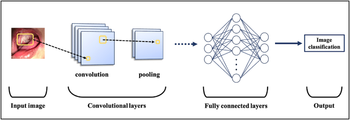
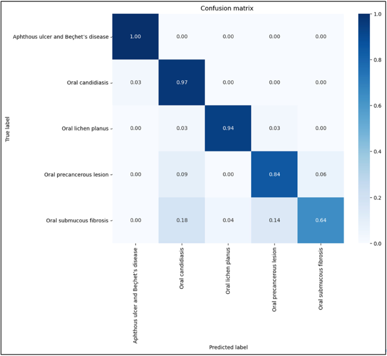
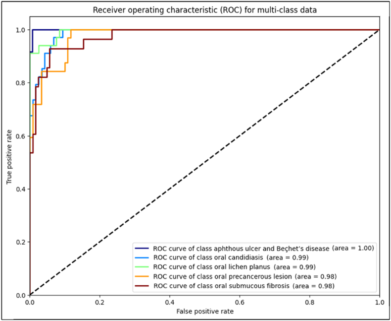
A set of clinical images were selected, including 506 images of five different diagnoses. The images were pre-processed and randomly divided into two sets for training and testing the CNN model. The model architecture was composed of convolutional layers, batch normalization layers, max pooling layers, the dropout layer and fully-connected layers. Evaluation metrics included weighted-precision, weighted-recall, weighted-F1 score, average specificity, Cohen’s Kappa coefficient, normalized confusion matrix and AUC.
Results
The overall performance for the image classification showed a weighted-precision of 88.8%, a weighted-recall of 88.2%, a weighted-F1 score of 0.878, an average pecificity of 97.0%, a Kappa coefficient of 0.851, and an average AUC of 0.985.
Conclusion
The model achieved a decent classification performance (overall AUC=0.985), showing the capacity to discern between benign and malignant potential lesions, and laid the foundation of a novel tool that can help clinical differential diagnosis of oral mucosal lesions. The main challenges were the small and imbalanced dataset. Enlarging the minority classes, incorporating more oral mucosal lesion diagnoses, employing transfer learning and cross-validation might be included in future works to optimize the image classification model.
期刊介绍:
he Journal of Dental Sciences (JDS), published quarterly, is the official and open access publication of the Association for Dental Sciences of the Republic of China (ADS-ROC). The precedent journal of the JDS is the Chinese Dental Journal (CDJ) which had already been covered by MEDLINE in 1988. As the CDJ continued to prove its importance in the region, the ADS-ROC decided to move to the international community by publishing an English journal. Hence, the birth of the JDS in 2006. The JDS is indexed in the SCI Expanded since 2008. It is also indexed in Scopus, and EMCare, ScienceDirect, SIIC Data Bases.
The topics covered by the JDS include all fields of basic and clinical dentistry. Some manuscripts focusing on the study of certain endemic diseases such as dental caries and periodontal diseases in particular regions of any country as well as oral pre-cancers, oral cancers, and oral submucous fibrosis related to betel nut chewing habit are also considered for publication. Besides, the JDS also publishes articles about the efficacy of a new treatment modality on oral verrucous hyperplasia or early oral squamous cell carcinoma.


 分享
分享
 求助内容:
求助内容: 应助结果提醒方式:
 扫码关注我们
