{"title":"Biphasic glucose-stimulated insulin secretion over decades: a journey from measurements and modeling to mechanistic insights.","authors":"Xiaohong Peng, Kai Wang, Liangyi Chen","doi":"10.1093/lifemeta/loae038","DOIUrl":null,"url":null,"abstract":"<p><p>Glucose-stimulated insulin release from pancreatic β-cells is critical for maintaining blood glucose homeostasis. An abrupt increase in blood glucose concentration evokes a rapid and transient rise in insulin secretion followed by a prolonged, slower phase. A diminished first phase is one of the earliest indicators of β-cell dysfunction in individuals predisposed to develop type 2 diabetes. Consequently, researchers have explored the underlying mechanisms for decades, starting with plasma insulin measurements under physiological conditions and advancing to single-vesicle exocytosis measurements in individual β-cells combined with molecular manipulations. Based on a chain of evidence gathered from genetic manipulation to <i>in vivo</i> mouse phenotyping, a widely accepted theory posits that distinct functional insulin vesicle pools in β-cells regulate biphasic glucose-stimulated insulin secretion (GSIS) via activation of different metabolic signal pathways. Recently, we developed a high-resolution imaging technique to visualize single vesicle exocytosis from β-cells within an intact islet. Our findings reveal that β-cells within the islet exhibit heterogeneity in their secretory capabilities, which also differs from the heterogeneous Ca<sup>2+</sup> signals observed in islet β-cells in response to glucose stimulation. Most importantly, we demonstrate that biphasic GSIS emerges from the interactions among α-, β-, and δ-cells within the islet and is driven by a small subset of hypersecretory β-cells. Finally, we propose that a shift from reductionism to holism may be required to fully understand the etiology of complex diseases such as diabetes.</p>","PeriodicalId":74074,"journal":{"name":"Life metabolism","volume":"4 1","pages":"loae038"},"PeriodicalIF":3.4000,"publicationDate":"2024-11-19","publicationTypes":"Journal Article","fieldsOfStudy":null,"isOpenAccess":false,"openAccessPdf":"https://www.ncbi.nlm.nih.gov/pmc/articles/PMC11770817/pdf/","citationCount":"0","resultStr":null,"platform":"Semanticscholar","paperid":null,"PeriodicalName":"Life metabolism","FirstCategoryId":"1085","ListUrlMain":"https://doi.org/10.1093/lifemeta/loae038","RegionNum":0,"RegionCategory":null,"ArticlePicture":[],"TitleCN":null,"AbstractTextCN":null,"PMCID":null,"EPubDate":"2025/2/1 0:00:00","PubModel":"eCollection","JCR":"","JCRName":"","Score":null,"Total":0}
引用次数: 0
Abstract
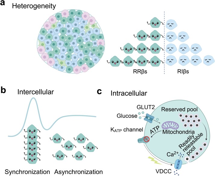
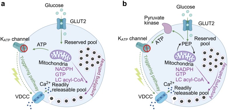
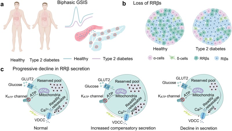
Glucose-stimulated insulin release from pancreatic β-cells is critical for maintaining blood glucose homeostasis. An abrupt increase in blood glucose concentration evokes a rapid and transient rise in insulin secretion followed by a prolonged, slower phase. A diminished first phase is one of the earliest indicators of β-cell dysfunction in individuals predisposed to develop type 2 diabetes. Consequently, researchers have explored the underlying mechanisms for decades, starting with plasma insulin measurements under physiological conditions and advancing to single-vesicle exocytosis measurements in individual β-cells combined with molecular manipulations. Based on a chain of evidence gathered from genetic manipulation to in vivo mouse phenotyping, a widely accepted theory posits that distinct functional insulin vesicle pools in β-cells regulate biphasic glucose-stimulated insulin secretion (GSIS) via activation of different metabolic signal pathways. Recently, we developed a high-resolution imaging technique to visualize single vesicle exocytosis from β-cells within an intact islet. Our findings reveal that β-cells within the islet exhibit heterogeneity in their secretory capabilities, which also differs from the heterogeneous Ca2+ signals observed in islet β-cells in response to glucose stimulation. Most importantly, we demonstrate that biphasic GSIS emerges from the interactions among α-, β-, and δ-cells within the islet and is driven by a small subset of hypersecretory β-cells. Finally, we propose that a shift from reductionism to holism may be required to fully understand the etiology of complex diseases such as diabetes.


 分享
分享

 求助内容:
求助内容: 应助结果提醒方式:
 扫码关注我们
