Tatiane Maria Rodrigues, Aline Rodrigues de Almeida, Juan de Nicolai, Igor Soares Dos Santos, Silvia Rodrigues Machado
{"title":"Interconnected idioblasts in <i>Peltaea polymorpha</i>: a novel component of the mucilage-secretory apparatus in Malvaceae.","authors":"Tatiane Maria Rodrigues, Aline Rodrigues de Almeida, Juan de Nicolai, Igor Soares Dos Santos, Silvia Rodrigues Machado","doi":"10.1093/aobpla/plae063","DOIUrl":null,"url":null,"abstract":"<p><p>The anatomical and cytological characteristics of the mucilage-secretory system have been widely studied in Malvaceae. However, conflicting information regarding the morphological nature of secretory structures exists, and some remain poorly understood. In this sense, some secretory structures in Malvaceae are not characterized as typical isolated idioblasts, canals, or cavities. Here, we describe a novel component of the mucilage-secretory apparatus in the Malvaceae family. Samples of the shoot apex, mature stem and fully expanded leaves were obtained from adult <i>Peltaea polymorpha,</i> which grow in the Cerrado (Brazilian savanna). The samples were processed using standard light and transmission electron microscopy methods. Mucilage cells occurred in the cortex and pith of petioles and stems, and in the midrib of leaves. These cells originate early in the stem apex from successive divisions of cells of the fundamental meristem, resulting in a row of interconnected secretory cells enveloped by a sheath of parenchyma cells devoid of secretory activity. Mucilage is stored in both protoplast and apoplast. In the same row, some cells filled with mucilage become very swollen and compress the neighbouring idioblasts that become flattened. This phenomenon results in a sandwich panel structure consisting of the swollen transversal walls of adjacent cells. As the differentiation progresses, the transversal walls of the rowed mucilage cells became very swollen, multilayered, and porous. Cytoplasmic strands cross such transversal walls connecting rowed cells. Mucilage-secreting cells in <i>P. polymorpha</i> are interconnected idioblasts and represent a novel component of the mucilage-secretory apparatus in Malvaceae. These findings open new avenues for understanding the structure and dynamics of mucilage-secreting cells from a functional perspective.</p>","PeriodicalId":48955,"journal":{"name":"AoB Plants","volume":"17 1","pages":"plae063"},"PeriodicalIF":2.4000,"publicationDate":"2025-01-10","publicationTypes":"Journal Article","fieldsOfStudy":null,"isOpenAccess":false,"openAccessPdf":"https://www.ncbi.nlm.nih.gov/pmc/articles/PMC11773387/pdf/","citationCount":"0","resultStr":null,"platform":"Semanticscholar","paperid":null,"PeriodicalName":"AoB Plants","FirstCategoryId":"99","ListUrlMain":"https://doi.org/10.1093/aobpla/plae063","RegionNum":3,"RegionCategory":"生物学","ArticlePicture":[],"TitleCN":null,"AbstractTextCN":null,"PMCID":null,"EPubDate":"2025/1/1 0:00:00","PubModel":"eCollection","JCR":"Q2","JCRName":"ECOLOGY","Score":null,"Total":0}
引用次数: 0
Abstract
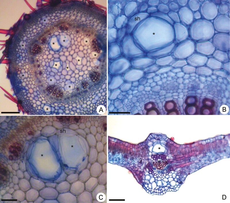
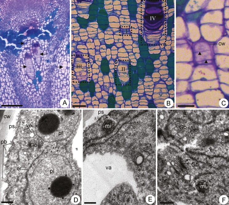
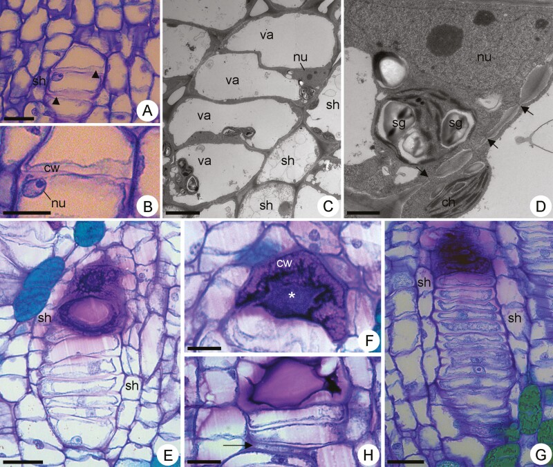
The anatomical and cytological characteristics of the mucilage-secretory system have been widely studied in Malvaceae. However, conflicting information regarding the morphological nature of secretory structures exists, and some remain poorly understood. In this sense, some secretory structures in Malvaceae are not characterized as typical isolated idioblasts, canals, or cavities. Here, we describe a novel component of the mucilage-secretory apparatus in the Malvaceae family. Samples of the shoot apex, mature stem and fully expanded leaves were obtained from adult Peltaea polymorpha, which grow in the Cerrado (Brazilian savanna). The samples were processed using standard light and transmission electron microscopy methods. Mucilage cells occurred in the cortex and pith of petioles and stems, and in the midrib of leaves. These cells originate early in the stem apex from successive divisions of cells of the fundamental meristem, resulting in a row of interconnected secretory cells enveloped by a sheath of parenchyma cells devoid of secretory activity. Mucilage is stored in both protoplast and apoplast. In the same row, some cells filled with mucilage become very swollen and compress the neighbouring idioblasts that become flattened. This phenomenon results in a sandwich panel structure consisting of the swollen transversal walls of adjacent cells. As the differentiation progresses, the transversal walls of the rowed mucilage cells became very swollen, multilayered, and porous. Cytoplasmic strands cross such transversal walls connecting rowed cells. Mucilage-secreting cells in P. polymorpha are interconnected idioblasts and represent a novel component of the mucilage-secretory apparatus in Malvaceae. These findings open new avenues for understanding the structure and dynamics of mucilage-secreting cells from a functional perspective.
期刊介绍:
AoB PLANTS is an open-access, online journal that has been publishing peer-reviewed articles since 2010, with an emphasis on all aspects of environmental and evolutionary plant biology. Published by Oxford University Press, this journal is dedicated to rapid publication of research articles, reviews, commentaries and short communications. The taxonomic scope of the journal spans the full gamut of vascular and non-vascular plants, as well as other taxa that impact these organisms. AoB PLANTS provides a fast-track pathway for publishing high-quality research in an open-access environment, where papers are available online to anyone, anywhere free of charge.


 分享
分享
 求助内容:
求助内容: 应助结果提醒方式:
 扫码关注我们
