Sandeep Dwarkanth Pande, Pala Kalyani, S Nagendram, Ala Saleh Alluhaidan, G Harish Babu, Sk Hasane Ahammad, Vivek Kumar Pandey, G Sridevi, Abhinav Kumar, Ebenezer Bonyah
{"title":"Comparative analysis of the DCNN and HFCNN Based Computerized detection of liver cancer.","authors":"Sandeep Dwarkanth Pande, Pala Kalyani, S Nagendram, Ala Saleh Alluhaidan, G Harish Babu, Sk Hasane Ahammad, Vivek Kumar Pandey, G Sridevi, Abhinav Kumar, Ebenezer Bonyah","doi":"10.1186/s12880-025-01578-4","DOIUrl":null,"url":null,"abstract":"<p><p>Liver cancer detection is critically important in the discipline of biomedical image testing and diagnosis. Researchers have explored numerous machine learning (ML) techniques and deep learning (DL) approaches aimed at the automated recognition of liver disease by analysing computed tomography (CT) images. This study compares two frameworks, Deep Convolutional Neural Network (DCNN) and Hierarchical Fusion Convolutional Neural Networks (HFCNN), to assess their effectiveness in liver cancer segmentation. The contribution includes enhancing the edges and textures of CT images through filtering to achieve precise liver segmentation. Additionally, an existing DL framework was employed for liver cancer detection and segmentation. The strengths of this paper include a clear emphasis on the criticality of liver cancer detection in biomedical imaging and diagnostics. It also highlights the challenges associated with CT image detection and segmentation and provides a comprehensive summary of recent literature. However, certain difficulties arise during the detection process in CT images due to overlapping structures, such as bile ducts, blood vessels, image noise, textural changes, size and location variations, and inherent heterogeneity. These factors may lead to segmentation errors and subsequently different analyses. This research analysis compares two advanced methodologies, DCNN and HFCNN, for liver cancer detection. The evaluation of DCNN and HFCNN in liver cancer detection is conducted using multiple performance metrics, including precision, F1-score, recall, and accuracy. This comprehensive assessment provides a detailed evaluation of these models' effectiveness compared to other state-of-the-art methods in identifying liver cancer.</p>","PeriodicalId":9020,"journal":{"name":"BMC Medical Imaging","volume":"25 1","pages":"37"},"PeriodicalIF":3.9000,"publicationDate":"2025-02-03","publicationTypes":"Journal Article","fieldsOfStudy":null,"isOpenAccess":false,"openAccessPdf":"https://www.ncbi.nlm.nih.gov/pmc/articles/PMC11792691/pdf/","citationCount":"0","resultStr":null,"platform":"Semanticscholar","paperid":null,"PeriodicalName":"BMC Medical Imaging","FirstCategoryId":"3","ListUrlMain":"https://doi.org/10.1186/s12880-025-01578-4","RegionNum":3,"RegionCategory":"医学","ArticlePicture":[],"TitleCN":null,"AbstractTextCN":null,"PMCID":null,"EPubDate":"","PubModel":"","JCR":"Q2","JCRName":"RADIOLOGY, NUCLEAR MEDICINE & MEDICAL IMAGING","Score":null,"Total":0}
引用次数: 0
Abstract
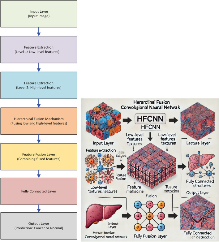
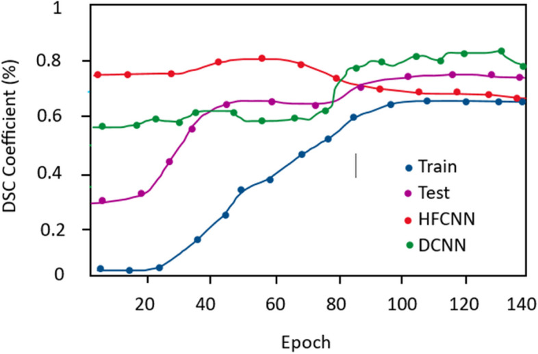
Liver cancer detection is critically important in the discipline of biomedical image testing and diagnosis. Researchers have explored numerous machine learning (ML) techniques and deep learning (DL) approaches aimed at the automated recognition of liver disease by analysing computed tomography (CT) images. This study compares two frameworks, Deep Convolutional Neural Network (DCNN) and Hierarchical Fusion Convolutional Neural Networks (HFCNN), to assess their effectiveness in liver cancer segmentation. The contribution includes enhancing the edges and textures of CT images through filtering to achieve precise liver segmentation. Additionally, an existing DL framework was employed for liver cancer detection and segmentation. The strengths of this paper include a clear emphasis on the criticality of liver cancer detection in biomedical imaging and diagnostics. It also highlights the challenges associated with CT image detection and segmentation and provides a comprehensive summary of recent literature. However, certain difficulties arise during the detection process in CT images due to overlapping structures, such as bile ducts, blood vessels, image noise, textural changes, size and location variations, and inherent heterogeneity. These factors may lead to segmentation errors and subsequently different analyses. This research analysis compares two advanced methodologies, DCNN and HFCNN, for liver cancer detection. The evaluation of DCNN and HFCNN in liver cancer detection is conducted using multiple performance metrics, including precision, F1-score, recall, and accuracy. This comprehensive assessment provides a detailed evaluation of these models' effectiveness compared to other state-of-the-art methods in identifying liver cancer.
期刊介绍:
BMC Medical Imaging is an open access journal publishing original peer-reviewed research articles in the development, evaluation, and use of imaging techniques and image processing tools to diagnose and manage disease.





 京公网安备 11010802042870号
京ICP备2023020795号-1
京公网安备 11010802042870号
京ICP备2023020795号-1

 分享
分享
 求助内容:
求助内容: 应助结果提醒方式:
 扫码关注我们
