Computed tomography and magnetic resonance imaging features of abdominal and pelvic leiomyomatosis peritonealis disseminata: A retrospective observational study.
Xin He, Xinxing Ma, Nan Jiang, Chunjiao Weng, Ling Yang
{"title":"Computed tomography and magnetic resonance imaging features of abdominal and pelvic leiomyomatosis peritonealis disseminata: A retrospective observational study.","authors":"Xin He, Xinxing Ma, Nan Jiang, Chunjiao Weng, Ling Yang","doi":"10.25259/JCIS_144_2024","DOIUrl":null,"url":null,"abstract":"<p><strong>Objectives: </strong>Leiomyomatosis peritonealis disseminata (LPD) is a rare and specific type of leiomyomatosis that is often misdiagnosed as malignant tumor with peritoneal metastasis, and accurate diagnosis is critical to treatment planning. The purpose of this study is to investigate the radiological features of LPD, analyze, and summarize its differential diagnosis and clinical features to improve the understanding of this rare disease.</p><p><strong>Material and methods: </strong>A retrospective analysis was conducted on clinical and radiological features from 10 patients with pathologically confirmed LPD between 2012 and 2024. The computed tomography (CT) and magnetic resonance imaging (MRI) findings were examined, focusing on parameters such as location, morphology, size, quantity, enhancement patterns, and their relationship with adjacent organs. In addition, the radiological features and the key points of differential diagnosis were summarized.</p><p><strong>Results: </strong>All the 10 LPD patients presented with multifocal lesions in the abdomen and pelvis, and 7 of them had a history of hysteromyoma surgery. The number of lesions was all ≥2, most of them were round or quasi-circular, with clear boundaries and smooth edges, did not invade the neighboring parenchymal organs, with a length of about 1.5~16.8 cm. The lesions were located in the pelvic cavity in 6 cases, the abdominal wall in 6 cases, the intestinal wall in 3 cases, the rectouterine pouch in 1 case, the omentum in 5 cases, the abdominal cavity in 1 case, and the mesentery in 1 case. There were 7 cases with minimal pelvic fluid and 1 case with liver spread. CT showed circular solid nodules with clear boundaries. The density of small lesions was homogeneous. Cystic changes were observed in some large lesions. On MRI, T1-weighted imaging showed hypo to isointense, T2-weighted imaging (T2WI) mostly showed hypointense, and T2WI in some large lesions showed slightly high signal intensity, diffusion-weighted imaging signal intensity was not higher than that of myometrium, apparent diffusion coefficient showed isointense, and solid components of the lesions were significantly more homogeneous enhanced after enhancement, and the enhancement degree was similar to that of normal myometrium.</p><p><strong>Conclusion: </strong>Although the imaging findings of LPD are similar to malignant tumors with peritoneal implantation and metastasis, they have certain characteristics, which are helpful for differential diagnosis combined with the clinical history of patients.</p>","PeriodicalId":15512,"journal":{"name":"Journal of Clinical Imaging Science","volume":"15 ","pages":"6"},"PeriodicalIF":1.3000,"publicationDate":"2025-01-29","publicationTypes":"Journal Article","fieldsOfStudy":null,"isOpenAccess":false,"openAccessPdf":"https://www.ncbi.nlm.nih.gov/pmc/articles/PMC11801444/pdf/","citationCount":"0","resultStr":null,"platform":"Semanticscholar","paperid":null,"PeriodicalName":"Journal of Clinical Imaging Science","FirstCategoryId":"1085","ListUrlMain":"https://doi.org/10.25259/JCIS_144_2024","RegionNum":0,"RegionCategory":null,"ArticlePicture":[],"TitleCN":null,"AbstractTextCN":null,"PMCID":null,"EPubDate":"2025/1/1 0:00:00","PubModel":"eCollection","JCR":"Q3","JCRName":"RADIOLOGY, NUCLEAR MEDICINE & MEDICAL IMAGING","Score":null,"Total":0}
引用次数: 0
Abstract
Objectives: Leiomyomatosis peritonealis disseminata (LPD) is a rare and specific type of leiomyomatosis that is often misdiagnosed as malignant tumor with peritoneal metastasis, and accurate diagnosis is critical to treatment planning. The purpose of this study is to investigate the radiological features of LPD, analyze, and summarize its differential diagnosis and clinical features to improve the understanding of this rare disease.
Material and methods: A retrospective analysis was conducted on clinical and radiological features from 10 patients with pathologically confirmed LPD between 2012 and 2024. The computed tomography (CT) and magnetic resonance imaging (MRI) findings were examined, focusing on parameters such as location, morphology, size, quantity, enhancement patterns, and their relationship with adjacent organs. In addition, the radiological features and the key points of differential diagnosis were summarized.
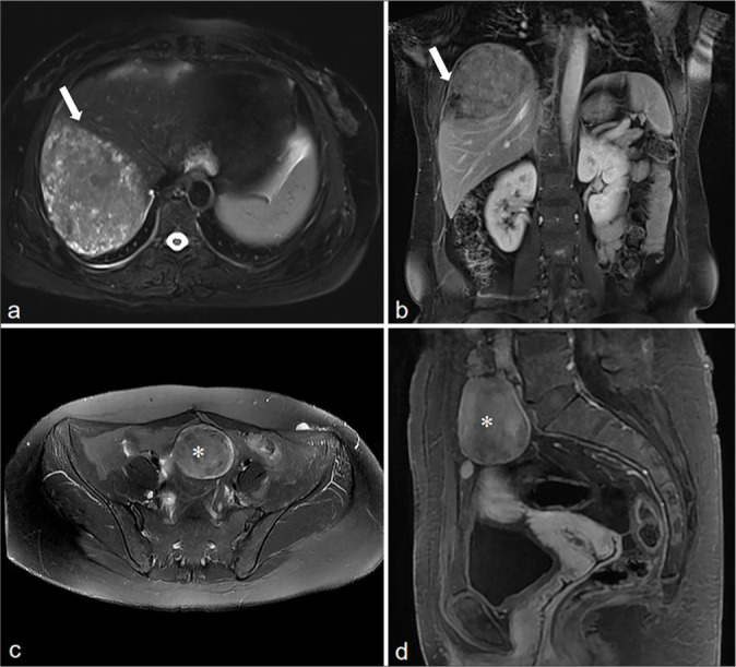
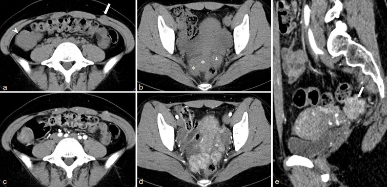
Results: All the 10 LPD patients presented with multifocal lesions in the abdomen and pelvis, and 7 of them had a history of hysteromyoma surgery. The number of lesions was all ≥2, most of them were round or quasi-circular, with clear boundaries and smooth edges, did not invade the neighboring parenchymal organs, with a length of about 1.5~16.8 cm. The lesions were located in the pelvic cavity in 6 cases, the abdominal wall in 6 cases, the intestinal wall in 3 cases, the rectouterine pouch in 1 case, the omentum in 5 cases, the abdominal cavity in 1 case, and the mesentery in 1 case. There were 7 cases with minimal pelvic fluid and 1 case with liver spread. CT showed circular solid nodules with clear boundaries. The density of small lesions was homogeneous. Cystic changes were observed in some large lesions. On MRI, T1-weighted imaging showed hypo to isointense, T2-weighted imaging (T2WI) mostly showed hypointense, and T2WI in some large lesions showed slightly high signal intensity, diffusion-weighted imaging signal intensity was not higher than that of myometrium, apparent diffusion coefficient showed isointense, and solid components of the lesions were significantly more homogeneous enhanced after enhancement, and the enhancement degree was similar to that of normal myometrium.
Conclusion: Although the imaging findings of LPD are similar to malignant tumors with peritoneal implantation and metastasis, they have certain characteristics, which are helpful for differential diagnosis combined with the clinical history of patients.
期刊介绍:
The Journal of Clinical Imaging Science (JCIS) is an open access peer-reviewed journal committed to publishing high-quality articles in the field of Imaging Science. The journal aims to present Imaging Science and relevant clinical information in an understandable and useful format. The journal is owned and published by the Scientific Scholar. Audience Our audience includes Radiologists, Researchers, Clinicians, medical professionals and students. Review process JCIS has a highly rigorous peer-review process that makes sure that manuscripts are scientifically accurate, relevant, novel and important. Authors disclose all conflicts, affiliations and financial associations such that the published content is not biased.


 分享
分享
 求助内容:
求助内容: 应助结果提醒方式:
 扫码关注我们
