Optimizing implant positioning in total hip arthroplasty via the direct anterior approach: The role and technique of conventional traction table and fluoroscopy.
{"title":"Optimizing implant positioning in total hip arthroplasty via the direct anterior approach: The role and technique of conventional traction table and fluoroscopy.","authors":"Seiya Ishii, Tomonori Baba, Koju Hayashi, Yasuhiro Homma, Osamu Muto, Muneaki Ishijima","doi":"10.1186/s42836-024-00293-9","DOIUrl":null,"url":null,"abstract":"<p><strong>Background: </strong>Precise implant positioning during total hip arthroplasty (THA) is an important factor influencing dislocation rate and long-term implant survival. Although a special carbon fiber traction table for THA improves the accuracy of implant positioning, it is too expensive. We aimed to report the accuracy of cup positioning and complication rate in patients undergoing THA via the direct anterior approach using a conventional noncarbon fiber traction table, which is generally used for osteosynthesis of femoral fractures.</p><p><strong>Methods: </strong>This retrospective study included 101 patients who received primary THA via the direct anterior approach using a conventional traction table with fluoroscopy between July 2022 and October 2024. Two observers evaluated radiological outcomes using postoperative anteroposterior X-rays. The intraclass correlation coefficients of cup positioning angles were calculated (inclination: 0.92, anteversion: 0.89 for intra-observer agreement; inclination: 0.91, anteversion: 0.85 for inter-observer agreement). Complications were defined as dislocation, periprosthetic fracture, ankle fracture, implant loosening, nerve injury, surgical site infection, deep vein thrombosis, and revision surgery for any reason.</p><p><strong>Results: </strong>Radiographic analysis showed an average cup inclination of 38.1° ± 4.1° (99.0% within Lewinnek's safe zone). The average cup anteversion was 12.0° ± 4.7° (97.0% within Lewinnek's safe zone). None of the patients experienced any complications.</p><p><strong>Conclusion: </strong>The use of a conventional traction table to perform THA using fluoroscopy may not interfere with precise cup positioning. This technique, which does not require a special carbon fiber traction table for THA, could be a feasible alternative for performing THA at general hospitals.</p>","PeriodicalId":52831,"journal":{"name":"Arthroplasty","volume":"7 1","pages":"10"},"PeriodicalIF":4.3000,"publicationDate":"2025-02-07","publicationTypes":"Journal Article","fieldsOfStudy":null,"isOpenAccess":false,"openAccessPdf":"https://www.ncbi.nlm.nih.gov/pmc/articles/PMC11804042/pdf/","citationCount":"0","resultStr":null,"platform":"Semanticscholar","paperid":null,"PeriodicalName":"Arthroplasty","FirstCategoryId":"3","ListUrlMain":"https://doi.org/10.1186/s42836-024-00293-9","RegionNum":4,"RegionCategory":"医学","ArticlePicture":[],"TitleCN":null,"AbstractTextCN":null,"PMCID":null,"EPubDate":"","PubModel":"","JCR":"Q2","JCRName":"ORTHOPEDICS","Score":null,"Total":0}
引用次数: 0
Abstract
Background: Precise implant positioning during total hip arthroplasty (THA) is an important factor influencing dislocation rate and long-term implant survival. Although a special carbon fiber traction table for THA improves the accuracy of implant positioning, it is too expensive. We aimed to report the accuracy of cup positioning and complication rate in patients undergoing THA via the direct anterior approach using a conventional noncarbon fiber traction table, which is generally used for osteosynthesis of femoral fractures.
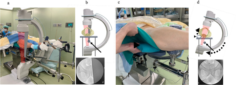
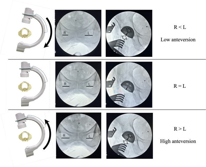
Methods: This retrospective study included 101 patients who received primary THA via the direct anterior approach using a conventional traction table with fluoroscopy between July 2022 and October 2024. Two observers evaluated radiological outcomes using postoperative anteroposterior X-rays. The intraclass correlation coefficients of cup positioning angles were calculated (inclination: 0.92, anteversion: 0.89 for intra-observer agreement; inclination: 0.91, anteversion: 0.85 for inter-observer agreement). Complications were defined as dislocation, periprosthetic fracture, ankle fracture, implant loosening, nerve injury, surgical site infection, deep vein thrombosis, and revision surgery for any reason.
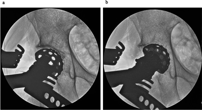
Results: Radiographic analysis showed an average cup inclination of 38.1° ± 4.1° (99.0% within Lewinnek's safe zone). The average cup anteversion was 12.0° ± 4.7° (97.0% within Lewinnek's safe zone). None of the patients experienced any complications.
Conclusion: The use of a conventional traction table to perform THA using fluoroscopy may not interfere with precise cup positioning. This technique, which does not require a special carbon fiber traction table for THA, could be a feasible alternative for performing THA at general hospitals.


 分享
分享
 求助内容:
求助内容: 应助结果提醒方式:
 扫码关注我们
