Bartosz Hanczaruk, Aleksandra Roszko, Kamil Kamiński, Marlena Tynecka
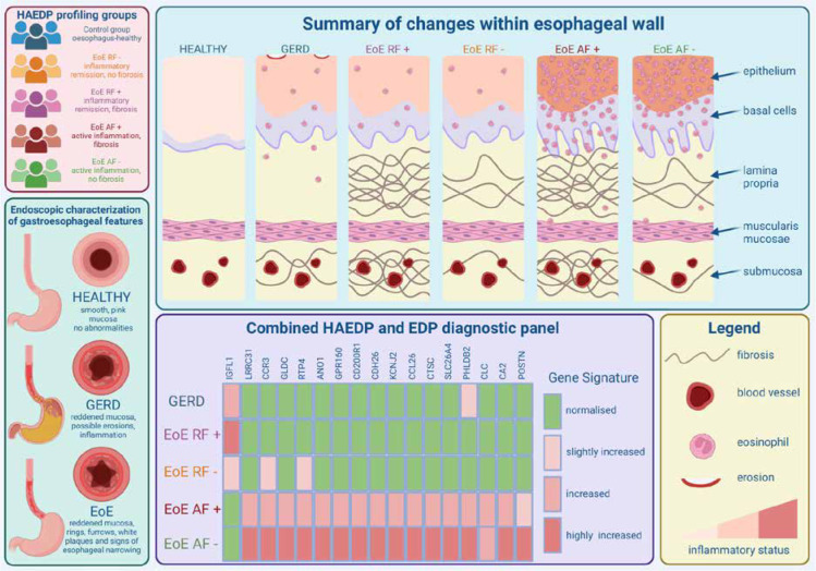
{"title":"Differential diagnosis of eosinophilic oesophagitis variants and biomarker identification: current progress.","authors":"Bartosz Hanczaruk, Aleksandra Roszko, Kamil Kamiński, Marlena Tynecka","doi":"10.5114/ceji.2024.143996","DOIUrl":null,"url":null,"abstract":"<p><p>Eosinophilic oesophagitis (EoE) is a disease characterized by dysregulated type 2 (T2) immune responses with enormous eosinophilic infiltration restricted to the oesophagus. Currently, the gold standard for EoE diagnosis involves identification of oesophageal dysfunction symptoms followed by the detection of at least 15 infiltrating eosinophils per high-power field in the oesophagus. Unfortunately, achieving 90% sensitivity in EoE histology-based diagnosis requires 5-6 biopsy samples to be collected from both the distal and proximal oesophagus, hindering precise diagnosis in routine clinical practice. Therefore, the development of novel diagnostic approaches differentiating EoE from other EoE-like diseases as well as identifying active and non-active forms of EoE is required. In line with the previously advanced EoE diagnostic panel (EDP), in a recent paper published in Gut (BMJ Journals), Gueguen et al. introduced a Histologically Active EoE Diagnostic Panel (HAEDP) effectively distinguishing patients with the active form of the disease from remission regardless of the fibrosis status and biopsy site. Here, we summarize recent findings and achievements in the development of the differential diagnosis of EoE based on the identification of unique deregulation in gene expression.</p>","PeriodicalId":9694,"journal":{"name":"Central European Journal of Immunology","volume":"49 4","pages":"438-441"},"PeriodicalIF":1.6000,"publicationDate":"2024-01-01","publicationTypes":"Journal Article","fieldsOfStudy":null,"isOpenAccess":false,"openAccessPdf":"https://www.ncbi.nlm.nih.gov/pmc/articles/PMC11811723/pdf/","citationCount":"0","resultStr":null,"platform":"Semanticscholar","paperid":null,"PeriodicalName":"Central European Journal of Immunology","FirstCategoryId":"3","ListUrlMain":"https://doi.org/10.5114/ceji.2024.143996","RegionNum":4,"RegionCategory":"医学","ArticlePicture":[],"TitleCN":null,"AbstractTextCN":null,"PMCID":null,"EPubDate":"2024/11/13 0:00:00","PubModel":"Epub","JCR":"Q4","JCRName":"IMMUNOLOGY","Score":null,"Total":0}
引用次数: 0
Abstract
Eosinophilic oesophagitis (EoE) is a disease characterized by dysregulated type 2 (T2) immune responses with enormous eosinophilic infiltration restricted to the oesophagus. Currently, the gold standard for EoE diagnosis involves identification of oesophageal dysfunction symptoms followed by the detection of at least 15 infiltrating eosinophils per high-power field in the oesophagus. Unfortunately, achieving 90% sensitivity in EoE histology-based diagnosis requires 5-6 biopsy samples to be collected from both the distal and proximal oesophagus, hindering precise diagnosis in routine clinical practice. Therefore, the development of novel diagnostic approaches differentiating EoE from other EoE-like diseases as well as identifying active and non-active forms of EoE is required. In line with the previously advanced EoE diagnostic panel (EDP), in a recent paper published in Gut (BMJ Journals), Gueguen et al. introduced a Histologically Active EoE Diagnostic Panel (HAEDP) effectively distinguishing patients with the active form of the disease from remission regardless of the fibrosis status and biopsy site. Here, we summarize recent findings and achievements in the development of the differential diagnosis of EoE based on the identification of unique deregulation in gene expression.


 分享
分享
 求助内容:
求助内容: 应助结果提醒方式:
 扫码关注我们
