Wilfried Mai, Silke Hecht, Matthew Paek, Shannon P Holmes, Hugo Dorez, Martin Blanchard, Jamil Nour Eddin
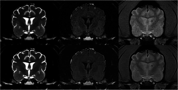
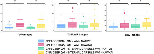
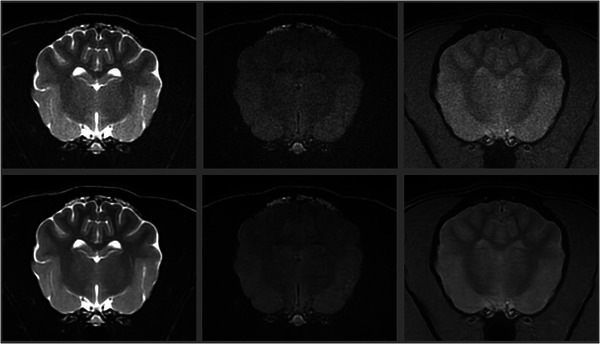
{"title":"A Veterinary DICOM-Based Deep Learning Denoising Algorithm Can Improve Subjective and Objective Brain MRI Image Quality.","authors":"Wilfried Mai, Silke Hecht, Matthew Paek, Shannon P Holmes, Hugo Dorez, Martin Blanchard, Jamil Nour Eddin","doi":"10.1111/vru.70015","DOIUrl":null,"url":null,"abstract":"<p><p>In this analytical cross-sectional method comparison study, we evaluated brain MR images in 30 dogs and cats with and without using a DICOM-based deep-learning (DL) denoising algorithm developed specifically for veterinary patients. Quantitative comparison was performed by measuring signal-to-noise (SNR) and contrast-to-noise ratios (CNR) on the same T2-weighted (T2W), T2-FLAIR, and Gradient Echo (GRE) MR brain images in each patient (native images and after denoising) in identical regions of interest. Qualitative comparisons were then conducted: three experienced veterinary radiologists independently evaluated each patient's T2W, T2-FLAIR, and GRE image series. Native and denoised images were evaluated separately, with observers blinded to the type of images they were assessing. For each image type (native and denoised) and pulse sequence type image, they assigned a subjective grade of coarseness, contrast, and overall quality. For all image series tested (T2W, T2-FLAIR, and GRE), the SNRs of cortical gray matter, subcortical white matter, deep gray matter, and internal capsule were statistically significantly higher on images treated with DL denoising algorithm than native images. Similarly, for all image series types tested, the CNRs between cortical gray and white matter and between deep gray matter and internal capsule were significantly higher on DL algorithm-treated images than native images. The qualitative analysis confirmed these results, with generally better coarseness, contrast, and overall quality scores for the images treated with the DL denoising algorithm. In this study, this DICOM-based DL denoising algorithm reduced noise in 1.5T MRI canine and feline brain images, and radiologists' perceived image quality improved.</p>","PeriodicalId":23581,"journal":{"name":"Veterinary Radiology & Ultrasound","volume":"66 2","pages":"e70015"},"PeriodicalIF":1.5000,"publicationDate":"2025-03-01","publicationTypes":"Journal Article","fieldsOfStudy":null,"isOpenAccess":false,"openAccessPdf":"https://www.ncbi.nlm.nih.gov/pmc/articles/PMC11822732/pdf/","citationCount":"0","resultStr":null,"platform":"Semanticscholar","paperid":null,"PeriodicalName":"Veterinary Radiology & Ultrasound","FirstCategoryId":"97","ListUrlMain":"https://doi.org/10.1111/vru.70015","RegionNum":2,"RegionCategory":"农林科学","ArticlePicture":[],"TitleCN":null,"AbstractTextCN":null,"PMCID":null,"EPubDate":"","PubModel":"","JCR":"Q2","JCRName":"VETERINARY SCIENCES","Score":null,"Total":0}
引用次数: 0
Abstract
In this analytical cross-sectional method comparison study, we evaluated brain MR images in 30 dogs and cats with and without using a DICOM-based deep-learning (DL) denoising algorithm developed specifically for veterinary patients. Quantitative comparison was performed by measuring signal-to-noise (SNR) and contrast-to-noise ratios (CNR) on the same T2-weighted (T2W), T2-FLAIR, and Gradient Echo (GRE) MR brain images in each patient (native images and after denoising) in identical regions of interest. Qualitative comparisons were then conducted: three experienced veterinary radiologists independently evaluated each patient's T2W, T2-FLAIR, and GRE image series. Native and denoised images were evaluated separately, with observers blinded to the type of images they were assessing. For each image type (native and denoised) and pulse sequence type image, they assigned a subjective grade of coarseness, contrast, and overall quality. For all image series tested (T2W, T2-FLAIR, and GRE), the SNRs of cortical gray matter, subcortical white matter, deep gray matter, and internal capsule were statistically significantly higher on images treated with DL denoising algorithm than native images. Similarly, for all image series types tested, the CNRs between cortical gray and white matter and between deep gray matter and internal capsule were significantly higher on DL algorithm-treated images than native images. The qualitative analysis confirmed these results, with generally better coarseness, contrast, and overall quality scores for the images treated with the DL denoising algorithm. In this study, this DICOM-based DL denoising algorithm reduced noise in 1.5T MRI canine and feline brain images, and radiologists' perceived image quality improved.
期刊介绍:
Veterinary Radiology & Ultrasound is a bimonthly, international, peer-reviewed, research journal devoted to the fields of veterinary diagnostic imaging and radiation oncology. Established in 1958, it is owned by the American College of Veterinary Radiology and is also the official journal for six affiliate veterinary organizations. Veterinary Radiology & Ultrasound is represented on the International Committee of Medical Journal Editors, World Association of Medical Editors, and Committee on Publication Ethics.
The mission of Veterinary Radiology & Ultrasound is to serve as a leading resource for high quality articles that advance scientific knowledge and standards of clinical practice in the areas of veterinary diagnostic radiology, computed tomography, magnetic resonance imaging, ultrasonography, nuclear imaging, radiation oncology, and interventional radiology. Manuscript types include original investigations, imaging diagnosis reports, review articles, editorials and letters to the Editor. Acceptance criteria include originality, significance, quality, reader interest, composition and adherence to author guidelines.


 分享
分享
 求助内容:
求助内容: 应助结果提醒方式:
 扫码关注我们
