{"title":"Unenhanced magnetic resonance imaging for the evaluation of sonographically indeterminate ovarian and adnexal masses.","authors":"Behnaz Moradi, Maryam Aghasi, Maryam Rahmani, Elham Sharifi, Mahrooz Malek, Fariba Yarandi, Masoumeh Banihashemian, Nadereh Behtash, Hamed Abdolghafoorian","doi":"10.1590/0100-3984.2024.0032","DOIUrl":null,"url":null,"abstract":"<p><strong>Objective: </strong>To investigate the accuracy of magnetic resonance imaging (MRI) in classifying sonographically indeterminate ovarian and adnexal masses.</p><p><strong>Materials and methods: </strong>This was a retrospective cross-sectional study of the unenhanced pelvic MRI scans of 243 patients with a collective total of 336 adnexal and ovarian masses.</p><p><strong>Results: </strong>Unenhanced MRI showed a sensitivity of 97.7%, a specificity of 86.4%, and an accuracy of 93.8%. The area under the ROC curve was 0.944 (95% CI: 0.913-0.974).</p><p><strong>Conclusion: </strong>Our results show that an unenhanced MRI protocol can be used to classify adnexal masses, especially in clinical settings in which the intravenous administration of gadolinium-based contrast is not safe and should be avoided.</p>","PeriodicalId":20842,"journal":{"name":"Radiologia Brasileira","volume":"58 ","pages":"e20240032"},"PeriodicalIF":0.0000,"publicationDate":"2025-02-07","publicationTypes":"Journal Article","fieldsOfStudy":null,"isOpenAccess":false,"openAccessPdf":"https://www.ncbi.nlm.nih.gov/pmc/articles/PMC11816912/pdf/","citationCount":"0","resultStr":null,"platform":"Semanticscholar","paperid":null,"PeriodicalName":"Radiologia Brasileira","FirstCategoryId":"1085","ListUrlMain":"https://doi.org/10.1590/0100-3984.2024.0032","RegionNum":0,"RegionCategory":null,"ArticlePicture":[],"TitleCN":null,"AbstractTextCN":null,"PMCID":null,"EPubDate":"2025/1/1 0:00:00","PubModel":"eCollection","JCR":"Q3","JCRName":"Medicine","Score":null,"Total":0}
引用次数: 0
Abstract
Objective: To investigate the accuracy of magnetic resonance imaging (MRI) in classifying sonographically indeterminate ovarian and adnexal masses.
Materials and methods: This was a retrospective cross-sectional study of the unenhanced pelvic MRI scans of 243 patients with a collective total of 336 adnexal and ovarian masses.
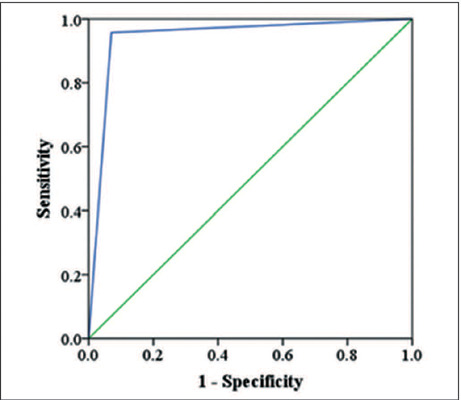
Results: Unenhanced MRI showed a sensitivity of 97.7%, a specificity of 86.4%, and an accuracy of 93.8%. The area under the ROC curve was 0.944 (95% CI: 0.913-0.974).
Conclusion: Our results show that an unenhanced MRI protocol can be used to classify adnexal masses, especially in clinical settings in which the intravenous administration of gadolinium-based contrast is not safe and should be avoided.


 分享
分享
 求助内容:
求助内容: 应助结果提醒方式:
 扫码关注我们
