Marta Dobek-Brylińska, Edyta Wlaźlak, Wiktor Wlaźlak, Jan Krakowiak, Andrzej Wróbel, Grzegorz Surkont
{"title":"The impact of pelvic floor contraction on urethral mobility and urogenital hiatus size in pelvic floor ultrasound.","authors":"Marta Dobek-Brylińska, Edyta Wlaźlak, Wiktor Wlaźlak, Jan Krakowiak, Andrzej Wróbel, Grzegorz Surkont","doi":"10.15557/JoU.2025.0005","DOIUrl":null,"url":null,"abstract":"<p><strong>Aim: </strong>The aim of the study was to assess the effect of pelvic floor contraction on urethral mobility and the size of the urogenital hiatus, as well as to compare two ultrasonographic approaches for the assessment of urethral mobility: transperineal with a transabdominal probe and transvestibular with a transvaginal transducer.</p><p><strong>Materials and methods: </strong>Modified Oxford Scale (MOS) was used for clinical evaluation of muscle contraction. The parameters obtained in both ultrasound approaches were assessed for all six Oxford grades. The values of ΔH, ΔD and vector, measured at rest and on pelvic floor muscle contraction, were used to evaluate urethral mobility parameters in both ultrasound methods. Patients with a history of urogynecological surgery, pelvic radiotherapy, significant pelvic prolapse (grade 2 or grater in at least one compartment), as well as those with unilateral or bilateral complete avulsion of the puborectalis muscle were excluded.</p><p><strong>Results: </strong>A total of 272 women were included in the analysis. A statistically significant correlation was found between the contraction force and urethral mobility parameters ΔH and vector-positive and ΔD-negative, obtained in both ultrasound approaches. However, no correlation was demonstrated between the contraction force and changes in the analyzed hiatal parameters. The Bland-Altman analysis showed a high agreement of both measurement methods.</p><p><strong>Conclusions: </strong>The force of pelvic floor muscle contraction, as measured with the Oxford Scale, correlated with urethral mobility in both ultrasound examinations. Assessment of urethral mobility using the three assessed parameters (ΔH, ΔD, vector) allows for the most comprehensive analysis. Only minor differences were found in the analyzed urethral mobility parameters between both ultrasonographic approaches. The impact of pelvic floor muscle contraction on the size of the urogenital hiatus was not confirmed.</p>","PeriodicalId":45612,"journal":{"name":"Journal of Ultrasonography","volume":"25 100","pages":"20250005"},"PeriodicalIF":1.5000,"publicationDate":"2025-02-19","publicationTypes":"Journal Article","fieldsOfStudy":null,"isOpenAccess":false,"openAccessPdf":"https://www.ncbi.nlm.nih.gov/pmc/articles/PMC11841739/pdf/","citationCount":"0","resultStr":null,"platform":"Semanticscholar","paperid":null,"PeriodicalName":"Journal of Ultrasonography","FirstCategoryId":"1085","ListUrlMain":"https://doi.org/10.15557/JoU.2025.0005","RegionNum":0,"RegionCategory":null,"ArticlePicture":[],"TitleCN":null,"AbstractTextCN":null,"PMCID":null,"EPubDate":"2025/1/1 0:00:00","PubModel":"eCollection","JCR":"Q3","JCRName":"RADIOLOGY, NUCLEAR MEDICINE & MEDICAL IMAGING","Score":null,"Total":0}
引用次数: 0
Abstract
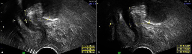
Aim: The aim of the study was to assess the effect of pelvic floor contraction on urethral mobility and the size of the urogenital hiatus, as well as to compare two ultrasonographic approaches for the assessment of urethral mobility: transperineal with a transabdominal probe and transvestibular with a transvaginal transducer.
Materials and methods: Modified Oxford Scale (MOS) was used for clinical evaluation of muscle contraction. The parameters obtained in both ultrasound approaches were assessed for all six Oxford grades. The values of ΔH, ΔD and vector, measured at rest and on pelvic floor muscle contraction, were used to evaluate urethral mobility parameters in both ultrasound methods. Patients with a history of urogynecological surgery, pelvic radiotherapy, significant pelvic prolapse (grade 2 or grater in at least one compartment), as well as those with unilateral or bilateral complete avulsion of the puborectalis muscle were excluded.
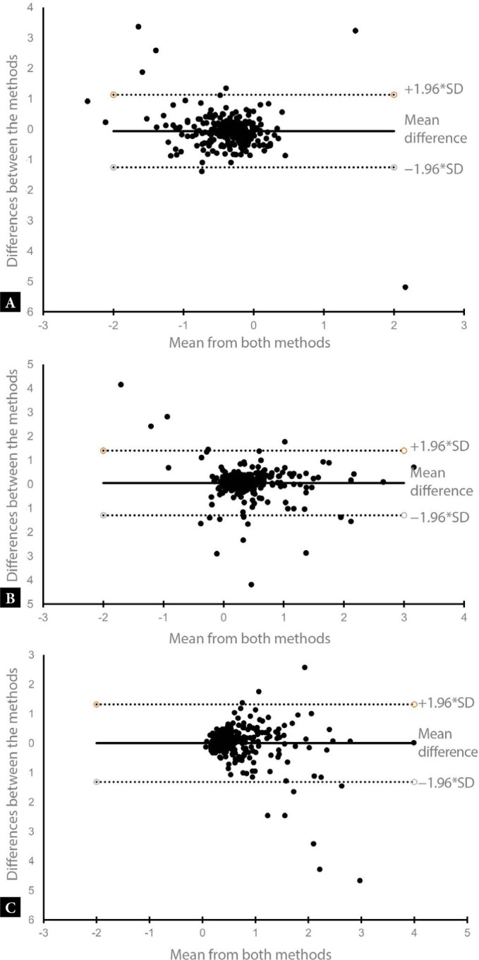
Results: A total of 272 women were included in the analysis. A statistically significant correlation was found between the contraction force and urethral mobility parameters ΔH and vector-positive and ΔD-negative, obtained in both ultrasound approaches. However, no correlation was demonstrated between the contraction force and changes in the analyzed hiatal parameters. The Bland-Altman analysis showed a high agreement of both measurement methods.
Conclusions: The force of pelvic floor muscle contraction, as measured with the Oxford Scale, correlated with urethral mobility in both ultrasound examinations. Assessment of urethral mobility using the three assessed parameters (ΔH, ΔD, vector) allows for the most comprehensive analysis. Only minor differences were found in the analyzed urethral mobility parameters between both ultrasonographic approaches. The impact of pelvic floor muscle contraction on the size of the urogenital hiatus was not confirmed.


 分享
分享
 求助内容:
求助内容: 应助结果提醒方式:
 扫码关注我们
