Marina París Ogáyar, Zeineb Ayed, Veronique Josserand, Maxime Henry, Álvaro Artiga, Livia Didonè, Miriam Granado, Aida Serrano, Ana Espinosa, Xavier Le Guével, Daniel Jaque
{"title":"Luminescence Fingerprint of Intracellular NIR-II Gold Nanocluster Transformation: Implications for Sensing and Imaging","authors":"Marina París Ogáyar, Zeineb Ayed, Veronique Josserand, Maxime Henry, Álvaro Artiga, Livia Didonè, Miriam Granado, Aida Serrano, Ana Espinosa, Xavier Le Guével, Daniel Jaque","doi":"10.1021/acsnano.4c13955","DOIUrl":null,"url":null,"abstract":"Gold nanoclusters emitting in the second biological window (NIR-II-AuNCs) have gained significant interest for their potential in deep-tissue bioimaging and biosensing applications due to the partial transparency and reduced autofluorescence of tissues in this spectral range. However, the limited understanding of how the biological environment affects their luminescent properties might hinder their use in bioimaging and biosensing. In this study, we investigated the emission properties of NIR-II-AuNCs when interacting and internalizing into live cells including macrophages, fibroblasts, and cancer cell lines, revealing substantial alterations in their luminescence. A systematic comparison between control and in vitro experiments concluded that the disruption of surface ligands is the main factor responsible for these alterations. NIR-II-AuNCs within cellular environments may also be influenced by other interactions, including aggregation or complexation with proteins. Furthermore, we also corroborated these spectroscopic modifications at the in vivo level, providing additional evidence of the environmental sensitivity of NIR-II-AuNCs. The results obtained in this study contribute to a deeper understanding of the luminescence mechanisms of NIR-II-AuNCs in biological environments in cells and in living tissues and are crucial for their optimization as reliable tools in biological environment for in vitro and in vivo imaging and diagnostics.","PeriodicalId":21,"journal":{"name":"ACS Nano","volume":"65 1","pages":""},"PeriodicalIF":16.0000,"publicationDate":"2025-02-24","publicationTypes":"Journal Article","fieldsOfStudy":null,"isOpenAccess":false,"openAccessPdf":"","citationCount":"0","resultStr":null,"platform":"Semanticscholar","paperid":null,"PeriodicalName":"ACS Nano","FirstCategoryId":"88","ListUrlMain":"https://doi.org/10.1021/acsnano.4c13955","RegionNum":1,"RegionCategory":"材料科学","ArticlePicture":[],"TitleCN":null,"AbstractTextCN":null,"PMCID":null,"EPubDate":"","PubModel":"","JCR":"Q1","JCRName":"CHEMISTRY, MULTIDISCIPLINARY","Score":null,"Total":0}
引用次数: 0
Abstract
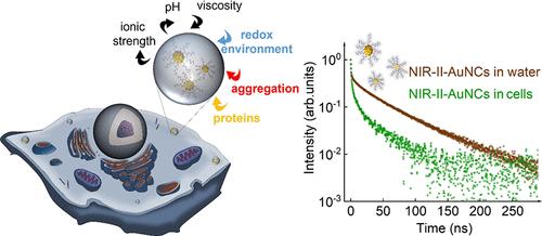
Gold nanoclusters emitting in the second biological window (NIR-II-AuNCs) have gained significant interest for their potential in deep-tissue bioimaging and biosensing applications due to the partial transparency and reduced autofluorescence of tissues in this spectral range. However, the limited understanding of how the biological environment affects their luminescent properties might hinder their use in bioimaging and biosensing. In this study, we investigated the emission properties of NIR-II-AuNCs when interacting and internalizing into live cells including macrophages, fibroblasts, and cancer cell lines, revealing substantial alterations in their luminescence. A systematic comparison between control and in vitro experiments concluded that the disruption of surface ligands is the main factor responsible for these alterations. NIR-II-AuNCs within cellular environments may also be influenced by other interactions, including aggregation or complexation with proteins. Furthermore, we also corroborated these spectroscopic modifications at the in vivo level, providing additional evidence of the environmental sensitivity of NIR-II-AuNCs. The results obtained in this study contribute to a deeper understanding of the luminescence mechanisms of NIR-II-AuNCs in biological environments in cells and in living tissues and are crucial for their optimization as reliable tools in biological environment for in vitro and in vivo imaging and diagnostics.
在第二生物窗口(NIR-II-AuNCs)中发射的金纳米团簇因其在深层组织成像和生物传感应用中的潜力而获得了极大的兴趣,这是由于该光谱范围内组织的部分透明度和减少的自身荧光。然而,对生物环境如何影响其发光特性的有限理解可能会阻碍其在生物成像和生物传感中的应用。在这项研究中,我们研究了NIR-II-AuNCs在巨噬细胞、成纤维细胞和癌细胞系等活细胞中相互作用和内化时的发光特性,揭示了它们发光的实质性变化。对照和体外实验之间的系统比较得出结论,表面配体的破坏是导致这些变化的主要因素。细胞环境中的nir - ii - aunc也可能受到其他相互作用的影响,包括与蛋白质的聚集或络合。此外,我们还在体内水平上证实了这些光谱修饰,为NIR-II-AuNCs的环境敏感性提供了额外的证据。本研究获得的结果有助于更深入地了解NIR-II-AuNCs在细胞和活组织生物环境中的发光机制,并对其作为生物环境中体外和体内成像和诊断的可靠工具进行优化至关重要。
期刊介绍:
ACS Nano, published monthly, serves as an international forum for comprehensive articles on nanoscience and nanotechnology research at the intersections of chemistry, biology, materials science, physics, and engineering. The journal fosters communication among scientists in these communities, facilitating collaboration, new research opportunities, and advancements through discoveries. ACS Nano covers synthesis, assembly, characterization, theory, and simulation of nanostructures, nanobiotechnology, nanofabrication, methods and tools for nanoscience and nanotechnology, and self- and directed-assembly. Alongside original research articles, it offers thorough reviews, perspectives on cutting-edge research, and discussions envisioning the future of nanoscience and nanotechnology.


 分享
分享
 求助内容:
求助内容: 应助结果提醒方式:
 扫码关注我们
