Cytological Diagnosis by Fine-Needle Aspiration or Core Biopsy with Touch Preparation for Renal Cystic or Solid Lesions: A Single-Center Clinicopathological Analysis.
Jiayan Liu, Changwei Yang, Xiaohui Wu, Li Yang, Hong Xu
{"title":"Cytological Diagnosis by Fine-Needle Aspiration or Core Biopsy with Touch Preparation for Renal Cystic or Solid Lesions: A Single-Center Clinicopathological Analysis.","authors":"Jiayan Liu, Changwei Yang, Xiaohui Wu, Li Yang, Hong Xu","doi":"10.1159/000543822","DOIUrl":null,"url":null,"abstract":"<p><strong>Introduction: </strong>A retrospective study analyzed real-life data from a single-center cohort of renal fine-needle aspiration (FNA) and core biopsy (CB) with touch preparation (TP) procedures over an 18-year period and aimed to provide a comprehensive overview of the evaluation of cytological diagnostic performance, challenges, and accuracy concerning renal cystic, solid, and mixed cystic-solid lesions.</p><p><strong>Methods: </strong>All percutaneous ultrasound-guided FNA and CT-guided CB with TP performed at our institution from 2006 to 2024 were identified.</p><p><strong>Results: </strong>A total of 89 renal cytology procedures were performed during the study period. Sixty-two (69.7%) lesions displayed cystic radiological features, 20 (22.5%) lesions presented solid mass, and only 7 (7.8%) lesions exhibited mixed cystic-solid radiological features. Of the procedures performed, seventy-five (84.3%) were ultrasound-guided FNA biopsies, while 14 (15.7%) were CT-guided CB with TP. Of the 62 renal cystic lesions, 54 (87.1%) were simple renal cysts. Twelve (60%) in 20 solid lesions were malignant, mainly involving clear cell renal cell carcinoma (RCC), urothelial carcinoma, and collecting duct carcinoma. Cytological diagnoses of renal mixed cystic-solid lesions mainly involved tuberculosis and clear cell RCC. However, only 22 cases had corresponding histopathological specimens available for comparison. The concordance rate between cytological diagnoses and surgical pathology specimens for cystic, solid, and mixed cystic-solid renal lesions was 100%, 92.3%, and 80%, respectively.</p><p><strong>Conclusion: </strong>In our series, FNA or CB with TP demonstrates significant diagnostic utility in the evaluation of renal lesions. The diagnostic accuracy of FNA cytology for renal lesions has been enhanced through the application of immunocytochemical staining on cell blocks.</p>","PeriodicalId":6959,"journal":{"name":"Acta Cytologica","volume":" ","pages":"210-220"},"PeriodicalIF":1.7000,"publicationDate":"2025-01-01","publicationTypes":"Journal Article","fieldsOfStudy":null,"isOpenAccess":false,"openAccessPdf":"https://www.ncbi.nlm.nih.gov/pmc/articles/PMC12052385/pdf/","citationCount":"0","resultStr":null,"platform":"Semanticscholar","paperid":null,"PeriodicalName":"Acta Cytologica","FirstCategoryId":"3","ListUrlMain":"https://doi.org/10.1159/000543822","RegionNum":4,"RegionCategory":"医学","ArticlePicture":[],"TitleCN":null,"AbstractTextCN":null,"PMCID":null,"EPubDate":"2025/2/25 0:00:00","PubModel":"Epub","JCR":"Q3","JCRName":"PATHOLOGY","Score":null,"Total":0}
引用次数: 0
Abstract
Introduction: A retrospective study analyzed real-life data from a single-center cohort of renal fine-needle aspiration (FNA) and core biopsy (CB) with touch preparation (TP) procedures over an 18-year period and aimed to provide a comprehensive overview of the evaluation of cytological diagnostic performance, challenges, and accuracy concerning renal cystic, solid, and mixed cystic-solid lesions.
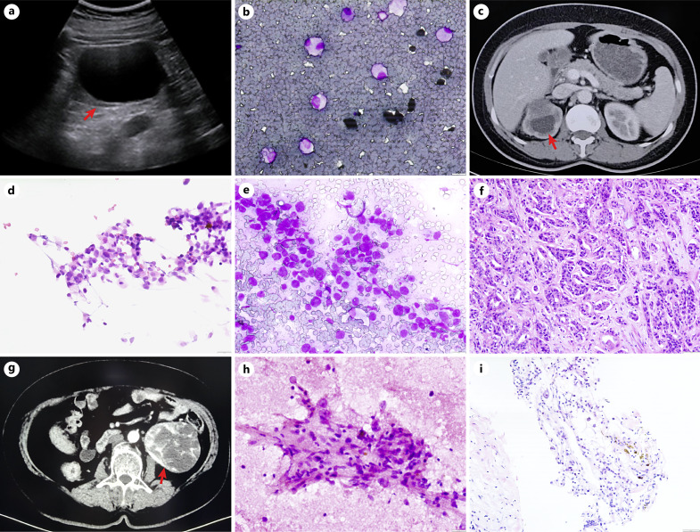
Methods: All percutaneous ultrasound-guided FNA and CT-guided CB with TP performed at our institution from 2006 to 2024 were identified.
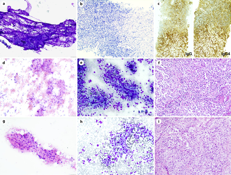
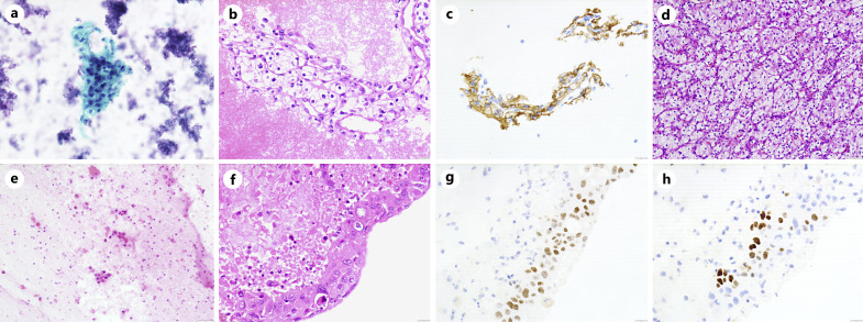
Results: A total of 89 renal cytology procedures were performed during the study period. Sixty-two (69.7%) lesions displayed cystic radiological features, 20 (22.5%) lesions presented solid mass, and only 7 (7.8%) lesions exhibited mixed cystic-solid radiological features. Of the procedures performed, seventy-five (84.3%) were ultrasound-guided FNA biopsies, while 14 (15.7%) were CT-guided CB with TP. Of the 62 renal cystic lesions, 54 (87.1%) were simple renal cysts. Twelve (60%) in 20 solid lesions were malignant, mainly involving clear cell renal cell carcinoma (RCC), urothelial carcinoma, and collecting duct carcinoma. Cytological diagnoses of renal mixed cystic-solid lesions mainly involved tuberculosis and clear cell RCC. However, only 22 cases had corresponding histopathological specimens available for comparison. The concordance rate between cytological diagnoses and surgical pathology specimens for cystic, solid, and mixed cystic-solid renal lesions was 100%, 92.3%, and 80%, respectively.
Conclusion: In our series, FNA or CB with TP demonstrates significant diagnostic utility in the evaluation of renal lesions. The diagnostic accuracy of FNA cytology for renal lesions has been enhanced through the application of immunocytochemical staining on cell blocks.
期刊介绍:
With articles offering an excellent balance between clinical cytology and cytopathology, ''Acta Cytologica'' fosters the understanding of the pathogenetic mechanisms behind cytomorphology and thus facilitates the translation of frontline research into clinical practice. As the official journal of the International Academy of Cytology and affiliated to over 50 national cytology societies around the world, ''Acta Cytologica'' evaluates new and existing diagnostic applications of scientific advances as well as their clinical correlations. Original papers, review articles, meta-analyses, novel insights from clinical practice, and letters to the editor cover topics from diagnostic cytopathology, gynecologic and non-gynecologic cytopathology to fine needle aspiration, molecular techniques and their diagnostic applications. As the perfect reference for practical use, ''Acta Cytologica'' addresses a multidisciplinary audience practicing clinical cytopathology, cell biology, oncology, interventional radiology, otorhinolaryngology, gastroenterology, urology, pulmonology and preventive medicine.


 分享
分享
 求助内容:
求助内容: 应助结果提醒方式:
 扫码关注我们
