Ali Işın, Özkan Köse, Emre Ak, Emel Emir Yetim, Can Çevikol, Tuba Melekoğlu
{"title":"The influence of the lower limb components on genu varum in football players: a full leg length magnetic resonance imaging study.","authors":"Ali Işın, Özkan Köse, Emre Ak, Emel Emir Yetim, Can Çevikol, Tuba Melekoğlu","doi":"10.1186/s13102-025-01075-9","DOIUrl":null,"url":null,"abstract":"<p><strong>Background: </strong>This study aimed to evaluate lower extremity alignment in football players with and without genu varum using magnetic resonance imaging (MRI) and to investigate the underlying mechanisms and contributing factors to malalignment.</p><p><strong>Methods: </strong>This prospective case-control study included 36 male football players aged 16-19 years, divided into two groups: 18 with genu varum and 18 controls with normal lower extremity alignment. Full-length lower extremity MRI was used to assess alignment parameters. The isokinetic strength of the concentric knee extensor-flexor and concentric hip abductor-adductor muscles was measured using an isokinetic dynamometer at angular velocities of 60°/sec and 180°/sec. Logistic regression was used to evaluate the risk factors for genu varum.</p><p><strong>Results: </strong>Genu varum group had a mean mechanical axis deviation (MAD) of 14 ± 5 mm (p < 0.001), with 11 players exceeding the clinical cutoff of 15 mm. Significant differences were observed in the lateral distal tibial angle (LDTA) (p = 0.014), lateral proximal femoral angle (LPFA) (p = 0.017), and medial distal femoral angle (mLDFA) (p = 0.002) between the groups. Muscle strength values were comparable between the groups, except for the hip adductor-abductor strength ratio at 60°/sec, which was significantly lower in the genu varum group (p = 0.008), while all other comparisons were non-significant (p > 0.05). The regression analysis demonstrated that the mechanisms responsible for varus alignment in football players differ between the dominant and non-dominant leg.</p><p><strong>Conclusions: </strong>The findings in this study suggest that the proximal tibial deformity is a key factor in malalignment among football players with genu varum. Differences in alignment were observed between dominant and non-dominant legs. Strength values were similar between players with and without varus alignment, except for the 60˚/sec angular velocity Add/Abd ratio.</p><p><strong>Clinical trial registration: </strong>NCT06606964 / 16.09.2024.</p><p><strong>Level of evidence: </strong>Level III.</p>","PeriodicalId":48585,"journal":{"name":"BMC Sports Science Medicine and Rehabilitation","volume":"17 1","pages":"25"},"PeriodicalIF":2.8000,"publicationDate":"2025-02-25","publicationTypes":"Journal Article","fieldsOfStudy":null,"isOpenAccess":false,"openAccessPdf":"https://www.ncbi.nlm.nih.gov/pmc/articles/PMC11863870/pdf/","citationCount":"0","resultStr":null,"platform":"Semanticscholar","paperid":null,"PeriodicalName":"BMC Sports Science Medicine and Rehabilitation","FirstCategoryId":"3","ListUrlMain":"https://doi.org/10.1186/s13102-025-01075-9","RegionNum":3,"RegionCategory":"医学","ArticlePicture":[],"TitleCN":null,"AbstractTextCN":null,"PMCID":null,"EPubDate":"","PubModel":"","JCR":"Q1","JCRName":"REHABILITATION","Score":null,"Total":0}
引用次数: 0
Abstract
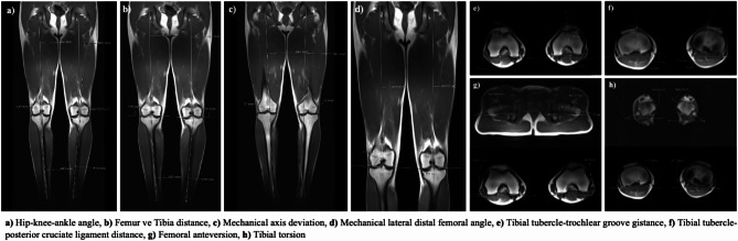
Background: This study aimed to evaluate lower extremity alignment in football players with and without genu varum using magnetic resonance imaging (MRI) and to investigate the underlying mechanisms and contributing factors to malalignment.
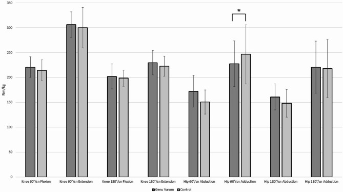
Methods: This prospective case-control study included 36 male football players aged 16-19 years, divided into two groups: 18 with genu varum and 18 controls with normal lower extremity alignment. Full-length lower extremity MRI was used to assess alignment parameters. The isokinetic strength of the concentric knee extensor-flexor and concentric hip abductor-adductor muscles was measured using an isokinetic dynamometer at angular velocities of 60°/sec and 180°/sec. Logistic regression was used to evaluate the risk factors for genu varum.
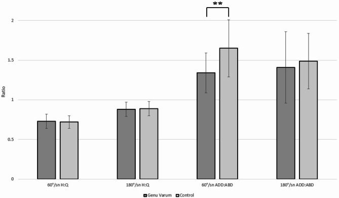
Results: Genu varum group had a mean mechanical axis deviation (MAD) of 14 ± 5 mm (p < 0.001), with 11 players exceeding the clinical cutoff of 15 mm. Significant differences were observed in the lateral distal tibial angle (LDTA) (p = 0.014), lateral proximal femoral angle (LPFA) (p = 0.017), and medial distal femoral angle (mLDFA) (p = 0.002) between the groups. Muscle strength values were comparable between the groups, except for the hip adductor-abductor strength ratio at 60°/sec, which was significantly lower in the genu varum group (p = 0.008), while all other comparisons were non-significant (p > 0.05). The regression analysis demonstrated that the mechanisms responsible for varus alignment in football players differ between the dominant and non-dominant leg.
Conclusions: The findings in this study suggest that the proximal tibial deformity is a key factor in malalignment among football players with genu varum. Differences in alignment were observed between dominant and non-dominant legs. Strength values were similar between players with and without varus alignment, except for the 60˚/sec angular velocity Add/Abd ratio.
期刊介绍:
BMC Sports Science, Medicine and Rehabilitation is an open access, peer reviewed journal that considers articles on all aspects of sports medicine and the exercise sciences, including rehabilitation, traumatology, cardiology, physiology, and nutrition.


 分享
分享
 求助内容:
求助内容: 应助结果提醒方式:
 扫码关注我们
