Left atrial myxoma associated with multivessel coronary embolism confirmed by cardiac magnetic resonance imaging: a case of myocardial infarction with non-obstructive coronary arteries.
Annalisa Pasquini, Andrea Caffè, Monica Filice, Rosa Lillo, Rocco Antonio Montone, Giovanni Alfonso Chiariello, Natalia Pavone, Marialisa Nesta, Maria Grandinetti, Piergiorgio Bruno, Francesco Burzotta, Massimo Massetti
{"title":"Left atrial myxoma associated with multivessel coronary embolism confirmed by cardiac magnetic resonance imaging: a case of myocardial infarction with non-obstructive coronary arteries.","authors":"Annalisa Pasquini, Andrea Caffè, Monica Filice, Rosa Lillo, Rocco Antonio Montone, Giovanni Alfonso Chiariello, Natalia Pavone, Marialisa Nesta, Maria Grandinetti, Piergiorgio Bruno, Francesco Burzotta, Massimo Massetti","doi":"10.1093/ehjimp/qyae124","DOIUrl":null,"url":null,"abstract":"<p><strong>Aims: </strong>Explore the diagnostic value of multimodal imaging in identifying considerably rare causes of myocardial infarction.</p><p><strong>Methods and results: </strong>We report a case of myocardial infarction with non-obstructive coronary arteries (MINOCA) probably due to coronary embolism associated with a left atrial myxoma. A 56-year-old male presented with non-ST-elevation myocardial infarction, with coronary angiography showing mild coronary atherosclerosis without significant epicardial stenosis. Transthoracic echocardiography and cardiac magnetic resonance imaging (CMR) revealed a large left atrial mass, suspected to be an atrial myxoma. CMR also showed an ischaemic pattern involving multiple coronary territories, suggesting coronary embolism as the cause of the MINOCA. The patient underwent successful surgical excision of the left atrial mass, and histopathology confirmed the diagnosis of cardiac myxoma.</p><p><strong>Conclusions: </strong>This case highlights the relevance of CMR in detecting ischaemic patterns in patients with a working diagnosis of MINOCA and underlines the diagnostic value of multimodal imaging in identifying considerably rare causes of myocardial infarction, such as myxoma-associated coronary embolism.</p>","PeriodicalId":94317,"journal":{"name":"European heart journal. Imaging methods and practice","volume":"2 3","pages":"qyae124"},"PeriodicalIF":0.0000,"publicationDate":"2024-12-02","publicationTypes":"Journal Article","fieldsOfStudy":null,"isOpenAccess":false,"openAccessPdf":"https://www.ncbi.nlm.nih.gov/pmc/articles/PMC11852255/pdf/","citationCount":"0","resultStr":null,"platform":"Semanticscholar","paperid":null,"PeriodicalName":"European heart journal. Imaging methods and practice","FirstCategoryId":"1085","ListUrlMain":"https://doi.org/10.1093/ehjimp/qyae124","RegionNum":0,"RegionCategory":null,"ArticlePicture":[],"TitleCN":null,"AbstractTextCN":null,"PMCID":null,"EPubDate":"2024/7/1 0:00:00","PubModel":"eCollection","JCR":"","JCRName":"","Score":null,"Total":0}
引用次数: 0
Abstract
Aims: Explore the diagnostic value of multimodal imaging in identifying considerably rare causes of myocardial infarction.
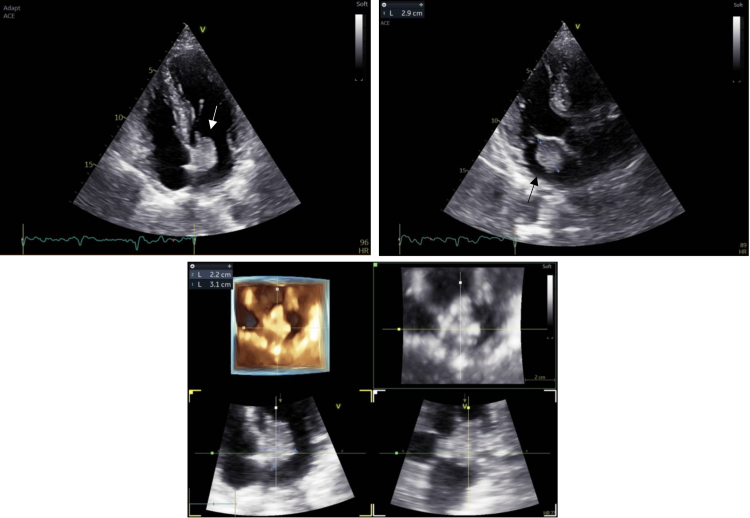
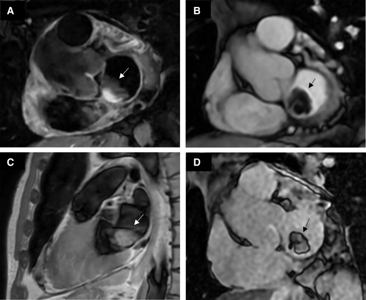
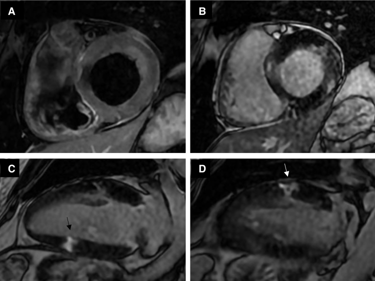
Methods and results: We report a case of myocardial infarction with non-obstructive coronary arteries (MINOCA) probably due to coronary embolism associated with a left atrial myxoma. A 56-year-old male presented with non-ST-elevation myocardial infarction, with coronary angiography showing mild coronary atherosclerosis without significant epicardial stenosis. Transthoracic echocardiography and cardiac magnetic resonance imaging (CMR) revealed a large left atrial mass, suspected to be an atrial myxoma. CMR also showed an ischaemic pattern involving multiple coronary territories, suggesting coronary embolism as the cause of the MINOCA. The patient underwent successful surgical excision of the left atrial mass, and histopathology confirmed the diagnosis of cardiac myxoma.
Conclusions: This case highlights the relevance of CMR in detecting ischaemic patterns in patients with a working diagnosis of MINOCA and underlines the diagnostic value of multimodal imaging in identifying considerably rare causes of myocardial infarction, such as myxoma-associated coronary embolism.


 分享
分享

 求助内容:
求助内容: 应助结果提醒方式:
 扫码关注我们
