{"title":"Maxillary Anterior Root Position/Angulation and Alveolar Bone Thickness in the Saudi Population: Implications for Implant Therapy.","authors":"Wesam Fathi, Kadi Alkheraije","doi":"10.1155/ijod/4469010","DOIUrl":null,"url":null,"abstract":"<p><p><b>Background:</b> Immediate implant placement has been considered a rapid and relatively efficient oral rehabilitation method that restores functional and esthetic demands. Understanding the anatomical tooth position and the natural dimensions of the alveolar ridge would facilitate proper treatment planning for immediate implants particularly in the esthetic zone. Therefore, the present study aims to use cone-beam computed tomography (CBCT) images to evaluate sagittal root position (SRP), tooth angulation within the alveolar ridge, and measuring labial and palatal alveolar bone thickness at maxillary anterior teeth. <b>Materials and Methods:</b> CBCT images of 102 Saudi adult subjects were used to evaluate the maxillary anterior teeth for three main parameters: SRP classification, root angulation in the alveolar bone, and labial and palatal bone thickness. <b>Results:</b> A total of 612 teeth were evaluated. Three hundred eighty-three teeth were classified as SRP Class I; majority of canines (75%), 46.08% of lateral incisors and 66.67% of central incisors. For SRP Class I, 46.5% of the teeth belong to females and 53.5% to males. In SRP Class II, 71.1% are female and 28.9% are male. SRP Class III has 33.3% females and 66.7% males. For SRP Class IV, 57.3% are female and 42.7% are male. Males have statistically significant thicker labial bone at canines and lateral incisors, at 2, 4, and 6 mm. However, males have statistically significant thicker bone at central incisors in palatal measurements and at the apex. <b>Conclusion:</b> There are significant differences in how canines, lateral, and central incisors are distributed across SRP classifications (<i>p</i> < 0.001). Canines show high frequency in Class I, but low in Class II. Lateral incisors have more even distribution between Class I and II. Central incisors follow similar pattern to canines with high Class I. The variations in SRP class and the gender differences in bone thickness identified in the current study confirm the necessity of personalized treatment plans to enhance immediate or even delayed implant placement success rates.</p>","PeriodicalId":13947,"journal":{"name":"International Journal of Dentistry","volume":"2025 ","pages":"4469010"},"PeriodicalIF":2.2000,"publicationDate":"2025-02-20","publicationTypes":"Journal Article","fieldsOfStudy":null,"isOpenAccess":false,"openAccessPdf":"https://www.ncbi.nlm.nih.gov/pmc/articles/PMC11867724/pdf/","citationCount":"0","resultStr":null,"platform":"Semanticscholar","paperid":null,"PeriodicalName":"International Journal of Dentistry","FirstCategoryId":"1085","ListUrlMain":"https://doi.org/10.1155/ijod/4469010","RegionNum":0,"RegionCategory":null,"ArticlePicture":[],"TitleCN":null,"AbstractTextCN":null,"PMCID":null,"EPubDate":"2025/1/1 0:00:00","PubModel":"eCollection","JCR":"Q2","JCRName":"DENTISTRY, ORAL SURGERY & MEDICINE","Score":null,"Total":0}
引用次数: 0
Abstract
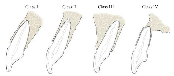
Background: Immediate implant placement has been considered a rapid and relatively efficient oral rehabilitation method that restores functional and esthetic demands. Understanding the anatomical tooth position and the natural dimensions of the alveolar ridge would facilitate proper treatment planning for immediate implants particularly in the esthetic zone. Therefore, the present study aims to use cone-beam computed tomography (CBCT) images to evaluate sagittal root position (SRP), tooth angulation within the alveolar ridge, and measuring labial and palatal alveolar bone thickness at maxillary anterior teeth. Materials and Methods: CBCT images of 102 Saudi adult subjects were used to evaluate the maxillary anterior teeth for three main parameters: SRP classification, root angulation in the alveolar bone, and labial and palatal bone thickness. Results: A total of 612 teeth were evaluated. Three hundred eighty-three teeth were classified as SRP Class I; majority of canines (75%), 46.08% of lateral incisors and 66.67% of central incisors. For SRP Class I, 46.5% of the teeth belong to females and 53.5% to males. In SRP Class II, 71.1% are female and 28.9% are male. SRP Class III has 33.3% females and 66.7% males. For SRP Class IV, 57.3% are female and 42.7% are male. Males have statistically significant thicker labial bone at canines and lateral incisors, at 2, 4, and 6 mm. However, males have statistically significant thicker bone at central incisors in palatal measurements and at the apex. Conclusion: There are significant differences in how canines, lateral, and central incisors are distributed across SRP classifications (p < 0.001). Canines show high frequency in Class I, but low in Class II. Lateral incisors have more even distribution between Class I and II. Central incisors follow similar pattern to canines with high Class I. The variations in SRP class and the gender differences in bone thickness identified in the current study confirm the necessity of personalized treatment plans to enhance immediate or even delayed implant placement success rates.


 分享
分享
 求助内容:
求助内容: 应助结果提醒方式:
 扫码关注我们
